2020.05.01.10
Files > Volume 5 > Vol 5 No 1 2020
REVISION/REVIEW
Extrapulmonary tuberculosis

Valarezo-Sevilla Diego1, Restrepo-Rodas Gabriela2, Sarzosa-Terán Vanessa3,
Available from: http://dx.doi.org/10.21931/RB/2020.05.01.10
SUMMARY
Extrapulmonary tuberculosis can be confused with other pathologies because of the variety of symptoms it generates according to the affected organ. So, extrapulmonary tuberculosis must always be taken into account by medical staff within the differential diagnosis.
In this paper, a review of the literature on extrapulmonary tuberculosis is carried out with emphasis on the most frequently affected organs.
Keywords. Extrapulmonary tuberculosis, HIV, social stigma, differential diagnosis
INTRODUCTION
Tuberculosis is an infectious disease caused by the Mycobacterium tuberculosis bacteria and is transmitted from person to person through the air. It is enough for a person to inhale a few bacilli to become infected. It is estimated that a quarter of the world's population has latent tuberculosis, this term applied to people infected by the bacillus but who have not yet become ill or can transmit the infection. Infected people have a lifetime risk of 5-15% of getting sick with tuberculosis; on the other hand, immunosuppressed people, for example, patients with HIV, malnutrition, diabetes, and smokers are at a much higher risk of becoming ill.1
Tuberculosis is one of the ten leading causes of mortality in the world. In 2017, 10 million people got infected with tuberculosis, and 1.6 million died from this disease (including 0.3 million people with HIV). Which is why tuberculosis is also considered as one of the leading causes of death among people with HIV. Within the pediatric population, it is estimated that in 2017 one million children got tuberculosis and 230,000 children died due to this cause (including children with HIV-associated tuberculosis). Tuberculosis is an infectious disease with a very high mortality rate; it causes around 4500 deaths every day. Communities bear the heaviest burden with socio-economic problems, those who work and live in high-risk environments and the poorest and most marginalized.2
The region of the Americas has significantly reduced new cases and deaths from tuberculosis in the last 25 years. Despite this, it is estimated that almost 270,000 people contracted the disease in 2015 and that nearly 50,000 do not know they have it yet. In the Americas, the population with a higher risk of infection are people with HIV, homeless, slum dwellers, deprived of liberty and people with addiction problems, communities that generally have limited access to health care and, if they do, they are not always diagnosed with tuberculosis when they suffer from it.3
The countries of the region are working to make the Americas the first area in the world to achieve the elimination of tuberculosis as a public health problem and currently, 15 countries have a low incidence of tuberculosis (Figure 1)4
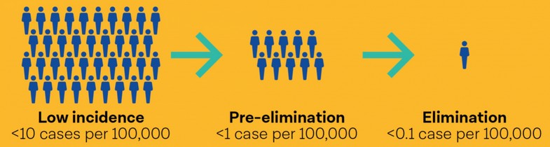
Figure 1. Steps towards tuberculosis elimination (taken from PAHO. Tuberculosis in the Americas 2018)
In 2015, the estimate by the World Health Organization for Ecuador was 8,400 new cases of tuberculosis (51.6 / 100 thousand inhabitants), including those with Tuberculosis / HIV coinfection. However, the National Health System (SNS) comprising the Comprehensive Public Health Network (RPIS) and the Complementary Network (RC) diagnosed and notified 5,215 cases (32.03 / 100 thousand inhabitants), fulfilling 62.08% of the estimated. Of the reported cases, 5,097 correspond to new and relapsed cases, and 118 previously treated cases (Ministry of Public Health 2017).5
Tuberculosis is described as the leading cause of death of infectious origin worldwide, therefore it is considered a global health problem; Taking this situation into account, we have reviewed the literature on extrapulmonary tuberculosis with emphasis on the most frequently affected organs.
Generalities of extrapulmonary tuberculosis
In general, the development of extrapulmonary tuberculosis is associated with immune deficiencies, genetic susceptibility, and unknown factors. Risk factors for extrapulmonary tuberculosis are immunosuppression, long-term illnesses such as HIV, children under 5, old age (over 60), female sex, race, and ethnicity (Asian or African origin). However, the large difference in the incidence of extrapulmonary tuberculosis between countries suggests that there are several associated factors such as; the increase of immigrant population that have a frequent delay in medical treatment, sub-registration, and genetic variations of the population and tuberculous mycobacteria (for example from Africa to Europe). Extrapulmonary tuberculosis has a high mortality rate, especially when considering the severity of some forms such as miliary or disseminated, and the prevalence of HIV.6
Early detection and treatment have been identified as essential elements in the Strategy of the World Health Organization (WHO) to end tuberculosis, a disease that remains a global health problem. Some of the challenges in the fight against tuberculosis are the inequality in access to medical care, resistance to multiple medications, co-infection with human immunodeficiency virus (HIV), the presence of a large group of latent infections and the delay in diagnosis.
Nonetheless, in high- and low-income countries, the delay in presentation, diagnosis and treatment of pulmonary tuberculosis is well documented. Which is why it is important to acknowledge that the rapid onset of tuberculosis treatment is not only crucial for the prognosis of the individual patient, since the delay in diagnosis has been associated with more serious clinical presentations, but also for the people who surround him because it carries a higher risk of transmission of Mycobacterium tuberculosis. Although the overall incidence of tuberculosis decreases in many regions, the increasing proportion of extrapulmonary tuberculosis is often neglected, consequently, a limited number of studies contain complete information on delays in the diagnosis of extrapulmonary tuberculosis.7
In some cases, the social stigma related to this highly transmissible pathology limits patients' access to health units. And in the cases that can overcome the social stigma, other factors such as physical, economic and geographic inaccessibility to health services (especially in rural areas) may play a role in late diagnosis and treatment.8
On average in the world, pulmonary tuberculosis represents 85% of clinical forms of tuberculosis, while extrapulmonary tuberculosis represents the remaining 15%. The most common types of extrapulmonary tuberculosis in adults include lymphatic, pleural, bone, meningeal, genitourinary and peritoneal tuberculosis. Nevertheless, the prevalence of extrapulmonary tuberculosis and its predominant forms varies from country to country. For example, Ethiopia has reported an incidence of 32%, which makes it the third country in the world, after India and Pakistan, with the highest rate of extrapulmonary tuberculosis; a number that has remained high over the years despite the country’s population size.
Global efforts to control tuberculosis have largely ignored extrapulmonary tuberculosis. This is because extrapulmonary tuberculosis is generally considered non-infectious and, as such, has no consequences on the global epidemic. However, recent data from northwestern England has shown that the prevalence of active tuberculosis disease among domestic contacts of extrapulmonary tuberculosis was high (440 per 100,000 contacts examined), which indicates that cases of extrapulmonary tuberculosis have a significant impact on tuberculosis transmission. It should also be contemplated that the slower annual decline rate of extrapulmonary tuberculosis compared to pulmonary tuberculosis could delay progress towards the END-TB targets set by the World Health Organization.9
The pediatric population, especially those under 3 years, have an incidence of up to 40% of developing tuberculosis after primary infection. Tuberculosis has a non-specific clinical presentation with a variety of imaging characteristics depending on the organs involved, which is why it often mimics other diseases. The most common site of tuberculosis in pediatric patients is the lungs (about 80%). The rest of the cases involve extrapulmonary sites: lymph nodes (67%), meninges (13%), pleura (6%), miliary dissemination (5%) and the musculoskeletal system (4%), with abdominal, renal and cutaneous involvement being less common [2]. Immunocompromised children, infants and adolescents have a higher risk of developing extrapulmonary tuberculosis. Pediatric extrapulmonary tuberculosis has an insidious onset without constitutional signs and symptoms in 72% of patients.10
The following can be considered as clinical clues to cause suspicion of extrapulmonary tuberculosis:11 Ascites with lymphocyte predominance and negative bacterial cultures; Chronic lymphadenopathy (especially cervical); CSF lymphocytic pleocytosis with elevated protein and low glucose; Differential diagnosis of Crohn's disease and amebiasis; Exudative pleural effusion with a predominance of lymphocytes, negative bacterial cultures and pleural thickening; HIV infection; Joint inflammation (monoarticular) with negative bacterial cultures; Persistent sterile pyuria; A country of origin that has endemic tuberculosis; Unexplained pericardial effusion, constrictive pericarditis, or pericardial calcification; Vertebral osteomyelitis that affects the thoracic spine.
Lymph node tuberculosis
Lymph node tuberculosis or tuberculous lymphadenitis is the most common form of extrapulmonary tuberculosis. According to its location, it is divided into intrathoracic and extrathoracic (also called peripheral lymphadenitis). Its most common location (the neck) has been known since ancient times as scrofula. The different routes of infection vary depending on which lymphatic group is affected. Cervical involvement is produced by direct contact of the bacilli with the Waldeyer ring and through it by dissemination and involvement of the adjacent ganglionic chains. It usually presents with unilateral, multiple and matt swelling of the neck in young adults, being a diagnosis and therapeutic challenge because it mimics other pathological processes, and it has an inconsistent physical performance and laboratory findings. The incidence of mycobacterial lymphadenitis has increased in parallel with the incidence of mycobacterial infection and HIV worldwide; so, it is important to differentiate tuberculous cervical lymphadenitis from non-tuberculous mycobacterial lymphadenitis, because their treatment protocols are different.12, 13
In the peripheral ganglionic form, the most frequent pathogenic mechanism is the reactivation of a primary pulmonary tuberculous infection, previously disseminated by the hematogenous route. Abdominal lymph node tuberculosis is also described by Mycobacterium bovis, secondary to ingestion of raw cow's milk, in less developed countries.12
Since tuberculosis is commonly considered a chronic lung disease, most studies of tuberculosis focus on the lungs, while lymph nodes are almost always represented only as antigen-presenting and immunological activation sites. Nonetheless, lymph nodes are among the most frequently infected sites of Mycobacterium tuberculosis, aside from the lungs. The effects of Mycobacterium tuberculosis infection and how lymph nodes respond to infection by this bacterium is currently unknown, as a consequence, many experimental studies have been conducted in macaques, which have found that general lymph nodes are not effective killers of Mycobacterium tuberculosis. In addition, the studies showed that the infection destroyed the lymph node structure and this, in turn, was associated with the increased bacterial load. After a brief course of antituberculous therapy, the reduction of the bacterial load was lower in the lymph nodes compared to the pulmonary granulomas. So, it was concluded that the lymph nodes, apart from being sites of antigen presentation and immune activation, can also be niches of growth and persistence of Mycobacterium tuberculosis.14
In lymphatic tuberculosis, the low culture rate of Mycobacterium tuberculosis forces the clinician to treat the disease without a drug sensitivity test of the evolving organism. This lack of evidence to base the choice of the drug leads to three important problems. First, there is an equally significant proportion of patients with Mycobacterium tuberculosis bacilli resistant to drugs in extrapulmonary tuberculosis as in pulmonary tuberculosis. Second, lymphatic tuberculosis usually responds slowly to tuberculosis treatment and the patient's condition may deteriorate paradoxically during therapy. Third, in extrapulmonary tuberculosis, there are no documented standard guidelines that stipulate the optimal duration of treatment or outcome like in pulmonary tuberculosis. Therefore, even though it is recommended that 6 months of standard tuberculosis treatment is adequate for most cases of lymphatic tuberculosis, many doctors fear the recurrence of tuberculosis in actual clinical practice, especially in cases treated without a drug resistance tests.15
In a study conducted in Denmark, tuberculous lymphadenitis was the most frequent manifestation of extrapulmonary tuberculosis; with a predominance in young immigrants and less frequently in vulnerable Danish patients. The majority of Mycobacterium tuberculosis isolated in this population harbored unique genotypes that suggest that tuberculous lymphadenitis could be associated with the reactivation of latent tuberculosis infection. Consequently, if this is really an association, future targeting of tuberculous lymphadenitis in risk groups could potentially reduce the number of cases in this country, with the vision of eliminating tuberculosis in the future.16
Extrapulmonary tuberculosis is most often diagnosed in peripheral lymph nodes. Due to the appearance of a lateral mass of the neck, the differential diagnosis may include neoplastic, infectious or immunological diseases, sarcoidosis, abscesses, tuberculosis, brucellosis, syphilis, toxoplasmosis, cat scratch disease and fungal infections. The presence of draining sinuses with purulent material suggests infectious and suppurative conditions such as pyogenic abscess, or other infectious processes.17
In another study conducted in the United Kingdom, it was shown that cervical tuberculous lymphadenitis was a condition that affected almost exclusively residents of the country that were born abroad in high-risk countries. The majority of patients were between 20 and 50 years old, generally in good health and had often resided in the United Kingdom for several years before the presentation. It was also found that the lymph nodes of the posterior triangle were more frequently involved and that constitutional symptoms occurred in a minority of patients.18
Extrapulmonary tuberculosis, although not as common as pulmonary tuberculosis, also exists in the United States. Their guidelines state that to reduce the differential diagnoses of a patient with isolated cervical or diffuse lymphadenopathy, an investigation into the country of origin and travel history should be made. Endoscopic ultrasound, fine needle aspiration, and fine needle biopsy are useful tools for the diagnosis of lymphatic tuberculosis, especially if the abdominal lymph nodes are the most accessible. The therapeutic approach is effective in most patients and surgical intervention can be avoided.19
The well-established treatment for patients with drug-susceptible tuberculosis lymphadenitis includes a 6-month regimen with isoniazid (INH), rifampicin (RIF), pyrazinamide (PZA) and ethambutol (EMB) during an intensive phase of 2 months, followed by a continuation phase of 4 months of treatment with INH and RIF. For HIV-TB coinfection, it has been suggested that the treatment protocol should include an antiretroviral therapy (ART) regimen. After 6 months of treatment, the reduction in lymph node size has been seen in less than 5 mm in 83% of patients. However, a paradoxical improvement reaction has also been observed in 20-30% of tuberculosis lymphadenitis. Surgical excision of ulcers and / or paranasal sinuses of the affected lymph nodes has been required to improve the effectiveness of the treatment when patients have little or no response to pharmacological treatment. Some variants have been reported in the duration of the continuation phase, because of cases with a poor response or because of episodes of drug resistance. As shown in a study, the 9-month regimen was recommended when treatment responses were delayed. It was reported that cure rates ranged from 89% to 94% when the duration of the regimen increased from 6 to 9 months, yet some patients may maintain persistent evidence of tuberculosis or develop relapses or recurrences after completing treatment.20
According to some findings, the diagnosis of tuberculous lymphadenitis requires a multifaceted approach that involves microbiology, pathology, radiology and other clinical specialties for the evaluation of suspicious cases. Laboratory findings should always be interpreted together with clinical pictures to consider a patient for an accurate diagnosis. The addition of culture for M. tuberculosis to the diagnostic algorithm together with fine-needle aspiration cytology and Ziehl-Neelsen staining helps to clarify the diagnostic dilemma with a high level of accuracy in cases of suspicion and may be useful in reducing tuberculosis burden. Still, more studies are needed to investigate the immune mechanism and its correlation with M. tuberculosis, so that patients that are negative for tuberculous lymphadenitis in fine-needle cytology but are positive for culture, can be diagnosed and treated with precision.21
Abdominal tuberculosis
The most common pathogenesis of both abdominal/peritoneal tuberculosis and tuberculous meningitis/encephalitis is the hematogenous spread of primary pulmonary tuberculosis. All of these diseases are forms of disseminated tuberculosis; however, only 15 to 25% of patients with abdominal tuberculosis and 40% of patients with tuberculosis meningitis have concomitant pulmonary tuberculosis. Extrapulmonary tuberculosis occurs most frequently months after pulmonary tuberculosis.22
Risk factors for abdominal/peritoneal tuberculosis specifically include alcoholism, liver disease, and cirrhosis, continuous ambulatory peritoneal dialysis for chronic renal failure, diabetes mellitus and Bacillus Calmette-Guérin therapy for superficial bladder carcinoma.22
Abdominal tuberculosis is an important form of extrapulmonary involvement, which is especially difficult to diagnose first due to the difficulty in tissue acquisition and second because it closely mimics certain conditions such as Crohn's disease and malignant neoplasia. In addition, the sequelae of intestinal tuberculosis, such as pain resulting from intestinal stenosis or peritoneal adhesions can be confused with the activity of the ongoing disease, which persuades doctors to prescribe a treatment of longer duration.23
Tuberculosis of the digestive system has many important relationships for its development. These relationships include some conditions, such as low socioeconomic status, immunosuppression status, as well as HIV infections, alcoholism or drug addiction. The symptoms presented in this disease are nonspecific, because the clinical picture is similar to that of other diseases, so the histopathological study remains the reference pattern for its diagnosis. In the literature, several mechanisms have been proposed by which tuberculosis spreads to the abdominal cavity, including the hematogenous, lymphatic or ingestion of mycobacteria.24
Pleural tuberculosis
Pleural tuberculosis should be suspected in any patient with unilateral pleural effusion of any size. It typically presents as an acute condition with fever (75%), chest pain with a pleuritic characteristic (50-75%) and non-productive cough (70-75%). More than two-thirds of patients are symptomatic for less than 1 month and the rest are symptomatic less than 1 week. Additional symptoms of presentation may be night sweats (50%), chills, weakness, dyspnea (50%), and weight loss (25-85%).25
In a study conducted in Italy, it was reported that 28% of cases of pleural tuberculosis were diagnosed as a result of thoracoscopy sampling. Spontaneous resolution of pleural tuberculosis is a well-known phenomenon, but 65% of patients can progress to pulmonary or extrapulmonary tuberculosis within 5 years. The usefulness of thoracoscopy for the diagnosis and treatment of pleural tuberculosis has already been previously reported, emphasizing that it is minimally invasive and can be performed under local anesthesia. Other diagnostic tests such as adenosine deaminase (ADA), interferon (IFN) -γ, interleukin-2, tumor necrosis factor-α, nucleic acid amplification tests (NAATs) and interferon-γ release assays are used to diagnose tuberculous pleural effusion but are only suggestive of the diagnosis, do not identify resistant mycobacteria and cannot guide treatment.26
You can also use a real-time polymerase chain reaction test for tuberculosis, which consists of a real-time amplification for the qualitative detection of the M. tuberculosis complex in biological materials (the result is obtained in an average of 3.5 days). The DNA is extracted from the samples; amplified and detected by fluorescent probes specific for M. tuberculosis.27
Currently, there is no diagnostic method with high sensitivity and specificity, low cost, ease and speed to perform it and that is widely available in areas where pleural tuberculosis prevails. Although it is usually observed in middle-aged men, with a time of evolution less than a month, such as a pleural exudate type exudate with a predominance of lymphocytes, the diagnosis of certainty has its limitations. Therefore, the clinical, epidemiology, Imaging studies, pathological anatomy and indirect laboratory tests, such as the determination of adenosine deaminase (ADA) levels, are a valuable contribution to the diagnosis.28
Despite the availability of countless diagnostic tests, and accurate test for pleural tuberculosis is still missing. The existing definition and criteria used for the classification of patients with pleural tuberculosis are not uniform; therefore, in the absence of a uniform case definition, it is a challenge to correctly evaluate and compare the performance of newly developed pleural tuberculosis diagnostic tests.29
Genitourinary tuberculosis
Renal involvement in tuberculosis may be part of a disseminated infection or a localized genitourinary disease. Urogenital tuberculosis accounts for 27% of extrapulmonary cases, even though renal involvement due to tuberculosis infection is underdiagnosed in most health centers. Most patients with renal tuberculosis have sterile pyuria (in the absence of common bacterial infection), which can be accompanied by microscopic hematuria.30
Genitourinary tuberculosis occurs due to hematogenous dissemination of tubercle bacilli, it can cause complications such as ureteral stenosis, chronic pyelonephritis and papillary necrosis, which can lead to impaired renal function. Tuberculous bacilli have a predilection for the upper and lower poles of the kidney and can form granulomas within the kidney that can remain indolent for many years. Image findings of renal tuberculosis result from the combination of papillary necrosis and parenchymal destruction.31
Since there is a high rate of false negatives (20%), a single urine culture-negative for mycobacteria does not mean the absence of genitourinary tuberculosis, so it is advisable to take at least 3 samples of the middle stream of the first-morning urine to be able to isolate the organism.32
For about 10% of patients with urogenital tuberculosis, the diagnosis is possible and is based on suggestive clinical, laboratory, and radiological findings, without microbiology or histological confirmation. The identification of the bacillus of tuberculosis in urine is achieved through Ziehl-Neelsen staining or by urine culture in Lowenstein-Jensen. The first one is fast, with 96.7% specificity but only 42.1 to 52.1% sensitivity. The culture is the gold standard for urogenital tuberculosis, with a tenderness that varies widely, from 10.7 to 90%, and the results may take 6 to 8 weeks to obtain. Since bacilluria is sporadic and weak, three to six urine samples are required.33
Renal tuberculosis with few clinical symptoms may be associated with several factors, one of them is the widespread use of broad-spectrum antibiotics; particularly fluoroquinolones. The symptoms may be masked by a nonspecific infection, calculus or tumor. It has also been described that doctors failed to identify or suspect renal tuberculosis in patients that did not show symptoms such as urinary frequency, urgency and dysuria, but showed back pain and hydronephrosis. Young doctors with little experience often settle for a diagnosis of kidney stones or tumor, while neglecting the possibility of renal tuberculosis.34
Fighting a global health problem
The diagnosis of extrapulmonary tuberculosis is a challenge and many patients start an empirical treatment against tuberculosis without a laboratory-confirmed diagnosis. Therefore, controlling the response to treatment is important to ensure a correct diagnosis and proper management of the disease, yet the definition of a satisfactory response to treatment in extrapulmonary tuberculosis remains unclear.35
It is recommended to use the same antibiotic regimen with a duration of 6 months. When there is the involvement of the central nervous system, the regimen should be prolonged to 12 months, and to in the case of tuberculous spondylitis with neurological involvement it should last 9 months. Mainly because in these patients, a shorter regimen has been associated with an increased risk of relapse. The treatment guideline is 2 months with rifampicin, isoniazid, pyrazinamide, and ethambutol, followed by 4 months of rifampicin and isoniazid. Once the sensitivity to first-line drugs is identified by antibiogram, ethambutol can be removed.36
Ensuring the proper management of household contacts of patients with tuberculosis is a logistical challenge. The cascade of attention for contact management presents many opportunities to get blocked: contacts may not be identified, evaluation procedures may not be available, therapy may not be prescribed, initiated or completed. Certain barriers have been consistently informed from various settings some of them is that contacts may face difficulties in traveling to health facilities for evaluation, that there are knowledge gaps between health workers and patients, and that there is low acceptability of taking medications among people who do not feel sick.37
National tuberculosis control programs in several countries have patient education programs that make emphasize only the cardinal symptoms of pulmonary tuberculosis.
Consequently, patients may not know about extrapulmonary tuberculosis, which is why it has been observed that many patients with extrapulmonary tuberculosis have advanced disease. Therefore, it is necessary to understand patients with extrapulmonary tuberculosis and their behavior to develop a strategy that improves diagnosis and treatment. The determination of extrapulmonary tuberculosis has always been a challenge for health care providers, and management generally requires more resources and more clinical experience than other diseases. In endemic tuberculosis sites with limited resources, the lack of availability of laboratory facilities in peripheral health centers often delays the diagnosis of extrapulmonary tuberculosis. Also, the increasing incidence rates and sparse literature on the various forms of extrapulmonary tuberculosis are grounds of concern that should be taken into account when designing significant diagnostic and treatment guidelines for national tuberculosis control programs.38
CONCLUSION
Extrapulmonary tuberculosis accounts for an essential percentage of patients with mycobacterial infection. Because many organs may be involved, extrapulmonary tuberculosis can present with a diversity of symptoms that can be confused with other pathologies. If the deceptive clinical presentation is associated with the lack of knowledge about this disease, an inevitable delay in both the diagnosis and the treatment results. Control programs must be improved by taking into account extrapulmonary tuberculosis as part of the public health problem.
REFERENCES
1. WHO. Tuberculosis. Geneva october 17, 2019. [Internet]. Available from:
https://www.who.int/es/news-room/fact-sheets/detail/tuberculosis
2. WHO. Nuevas recomendaciones de la OMS para acelerar los progresos en la lucha contra la tuberculosis. Geneva march 20, 2019. [Internet]. Available from:
https://www.who.int/es/news-room/detail/20-03-2019-new-who-recommendations-to-accelerate-progress-on-tb
3. PAHO/WHO. Tuberculosis: OPS/OMS llama a no dejar a nadie atrás. [Internet]. Available from:
https://www.paho.org/ecu/index.php?option=com_content&view=article&id=1882:tuberculosis-ops-oms-llama-a-no-dejar-a-nadie-atras&Itemid=360
4. PAHO. Tuberculosis en las Américas 2018. Washington, D.C. [Internet]. Available from: http://iris.paho.org/xmlui/bitstream/handle/123456789/49510/OPSCDE18036_spa?sequence=2&isAllowed=y
5. Alcívar-Solórzano LP, Arteaga-Intriago MA, Cando-Suviaga MA; Vinces-Sornoza TP, Macías-Alcívar EM, Cevallos-Garay WA. Factores que inciden para la presencia de tuberculosis. Dom. Cien. 2018; 4(4): 69-97. DOI: http://dx.doi.org/10.23857/dc.v4i4.824
6. Arnedo-Pena A, Romeu-Garcia MA, Meseguer-Ferrer N, Vivas-Fornas I, Vizcaino-Batllés A, Safont-Adsuara L, et al. Pulmonary versus extrapulmonary tuberculosis associated factors: A case-case study. Microbiology Insights 2019; 12: 1–10. DOI: 10.1177/1178636119840362
7. Mathiasen VD, Hansen AK, Eiset AH, Lillebaek T, Wejse C. Delays in the diagnosis and treatment of tuberculous lymphadenitis in low-incidence countries: a systematic review. Respiration. 2019. 9p. DOI: 10.1159/000499052
8. Adhikari S, Basnyat B. Extrapulmonary tuberculosis: a debilitating and often neglected public health problem. BMJ Case Rep 2018;11:e226098. DOI:10.1136/bcr-2018-226098
9. Mekonnen D, Derbie A, Abeje A, Shumet A, Nibret E, Biadglegne F, et al. Epidemiology of tuberculous lymphadenitis in Africa: a systematic review and meta-analysis. PLoS ONE. 2019; 14(4): e0215647. DOI: https://doi.org/10.1371/journal.pone.0215647
10. Kritsaneepaiboon S, Andres MM, Tatco VR, Lim CCQ, Concepcion NDP. Extrapulmonary involvement in pediatric tuberculosis. Pediatr Radiol 2017; 47:1249–59. DOI: 10.1007/s00247-017-3867-0
11. Golden MP, Vikram HR. Extrapulmonary tuberculosis: an overview. Am Fam Physician 2005; 72: 1761-8
12. Aliaga F, Rodríguez JC, Farga V. Reacciones paradójicas en el tratamiento de la tuberculosis ganglionar. Rev Chil Enferm Respir 2016; 32: 119-126
13. Cataño JC, Robledo J. Tuberculous lymphadenitis and parotitis. Microbiol Spectrum 2016; 4(6). DOI: 10.1128/microbiolspec.TNMI7-0008-2016
14. Ganchua SKC, Cadena AM, Maiello P, Gideon HP, Myers AJ, Junecko BF, et al. Lymph nodes are sites of prolonged bacterial persistence during Mycobacterium tuberculosis infection in macaques. PLoS Pathog 14(11):e1007337. DOI: https://doi.org/10.1371/journal.ppat.1007337
15. Ko Y, Kim C, Park YB, Mo EK, 1, Moon JW, Park S, et al. Clinical characteristics and treatment outcomes of definitive versus standard anti-tuberculosis therapy in patients with tuberculous lymphadenitis. J. Clin. Med. 2019; 8, 813; 10 p. DOI: http://dx.doi.org/10.3390/jcm8060813
16. Mathiasen VD, Eiset AH, Andersen PH, Wejse C, Lillebaek T. Epidemiology of tuberculous lymphadenitis in Denmark: a nationwide register-based study. PLoS ONE 2019; 14(8):e0221232. DOI: https://doi.org/10.1371/journal.pone.0221232
17. Michaelides SA , Bablekos GD , Michailidis AR, Gkioxari E, Vgenopoulou S, Chorti M. Left Lateral Cervical Mass with Draining Sinuses. Case Reports in Medicine. 2019; 6 pages
18. Moualed D, Robinson M, Qureishi A, Gurr P. Cervical tuberculous lymphadenitis: diagnosis and demographics, a five-year case series in the UK. Ann R Coll Surg Engl 2018; 100: 392–6. DOI: 10.1308/rcsann.2018.0021
19. Mehmood A, Ehsan A, Mukhtar M, Inayat F, Ullah W. Acute mesenteric tuberculous lymphadenitis: a comparative analysis of twenty-one cases. Cureus 2019; 11(4): e4454. 6p. DOI: 10.7759/cureus.4454
20. Qian X, Albers AE, Nguyen DTM, Dong Y, Zhang Y, Schreiber F, et al. Head and neck tuberculosis: literature review and meta-analysis. Tuberculosis. 2019. DOI: https://doi.org/10.1016/j.tube.2019.04.014
21. Kant K, Baveja CP, Sarkar J, Juya D. Microbiological evaluation of clinically suspected cases of tubercular lymphadenopathy by cytology, culture, and smear microscopy – A hospital-based study from Northern India. J Family Med Prim Care. 2019; 8(3):828–833. DOI: 10.4103/jfmpc.jfmpc_20_19
22. Cherabie J, Moore T. A Case of Extrapulmonary Tuberculosis. Two-ways. Kansas Journal of Medicine: 20-1
23. Mandavdhare HS, Singh H, Dutta U, Sharma V. A real-world experience with 6 months of antitubercular therapy in abdominal tuberculosis. JGH Open 2019; 3: 201–5. DOI: 10.1002/jgh3.12136
24. Gómez-Piña JJ. Tuberculosis peritoneal. Med Int Méx. 2018; 34(3):490-6. DOI: https://doi.org/10.24245/mim.v34i3.2171
25. Shaw JA, Irusen EM, Diacon AH, Koegelenberg CF. Pleural tuberculosis: A concise clinical review. Clin Respir J. 2018; 12(5):1779-86. DOI: 10.1111/crj.12900
26. Casalini AG, Mori PA, Majori M, Anghinolfi M, Silini EM, Gnetti L, et al. Pleural tuberculosis: medical thoracoscopy greatly increases the diagnostic accuracy. ERJ Open Res. 2018; 4(1):00046-2017. DOI: [https://doi.org/10.1183/23120541.00046-2017].
27.Casallas-Rivera MA, Bernal AMC, Giraldo-Cadavid LF, Prieto DE, Santander SP. Reacción en cadena de la polimerasa en tiempo real para el diagnóstico de tuberculosis pleural. Colomb Médica. 2017;48:7.
28. Golemba AS, Ferreyra FGE, Rovai GB, Achinelli FR. Tuberculosis pleural en un hospital del noreste argentino. MEDICINA (Buenos Aires) 2016; 76: 76-80
29. Tyagi S, Sharma N, Tyagi JS, Haldar S. Challenges in pleural tuberculosis diagnosis: existing reference standards and nucleic acid tests. Future Microbiol. 2017;12(13):1201-18. DOI: https://doi.org/10.2217/fmb-2017-0028
30. Daher EDF, Barros EJG, da Silva Junior GB. Renal Tuberculosis in the Modern Era. Am J Trop Med Hyg. 2013; 88(1):54-64. DOI:10.4269/ajtmh.2013.12-0413
31. Pinto DS, George A, Kumar N, Hoisala VR. A case report of renal papillary necrosis due to tuberculosis-CT urogram and static MR urogram findings. BJRcase Rep. 2017;3(2):20150438. DOI: https://doi.org/10.1259/bjrcr.20150438
32. Sourial MW, Brimo F, Horn R, Andonian S. Genitourinary tuberculosis in North America: A rare clinical entity. Can Urol Assoc J. 2015; 9(7-8):484. DOI: http://dx.doi.org/10.5489/cuaj.2643
33. Figueiredo AA, Lucon AM, Srougi M. Urogenital Tuberculosis. Microbiol Spectr 2017 [Internet]. [citado 28 de octubre de 2019];5(1). DOI: 10.1128/microbiolspec.TNMI7-0015-2016. Disponible en: http://www.asmscience.org/content/journal/microbiolspec/10.1128/microbiolspec.TNMI7-0015-2016
34. Wang J, Fan S, Xiao J, Liang C-Z. Renal tuberculosis tends to be low symptoms: how to improve the diagnosis and treatment of renal tuberculosis. Asian J Androl. 2016;18(1):145. DOI: 10.4103/1008-682X.150839
35. Jørstad MD, Dyrhol-Riise AM, Aßmus J, Marijani M, Sviland L, Mustafa T. Evaluation of treatment response in extrapulmonary tuberculosis in a low-resource setting. BMC Infectious Diseases 2019; 19:426; 9p. DOI: https://doi.org/10.1186/s12879-019-4034-z
36. Ramírez-Lapausa M, Menéndez-Saldaña A, Noguerado-Asensio A. Tuberculosis extrapulmonar, una revisión. Rev Esp Sanid Penit 2015; 17: 3-11
37. Yuen CM, Millones AK, Contreras CC, Lecca L, Becerra MC, Keshavjee S. Tuberculosis household accompaniment to improve the contact management cascade: a prospective cohort study. PLoS ONE. 2019; 14(5):e0217104. DOI: https://doi.org/10.1371/journal.pone.0217104
38. Purohit MR, Purohit R, Mustafa T. Patient health seeking and diagnostic delay in extrapulmonary tuberculosis: a hospital based study from central India. Tuberculosis Research and Treatment. 2019, 8 p. DOI: https://doi.org/10.1155/2019/4840561
Received: 2 noviembre 2019
Accepted: 15 enero 2020
Valarezo-Sevilla Diego1, Restrepo-Rodas Gabriela2, Sarzosa-Terán Vanessa3,
1 Especialista en Medicina Interna, Hospital General Ibarra
2 Estudiante de Medicina, Universidad Internacional del Ecuador
3, Especialista en Medicina Interna, Hospital Básico Antonio Ante
Corresponding author
Diego Valarezo Sevilla
ORCID: 0000-0001-9477-6261
Hospital General Ibarra, Ibarra - Ecuador
