2022.07.03.24
Files > Volume 7 > Vol 7 No 3 2022
Study of HLA-G gene polymorphism and serum level of soluble HLA-G in patients infected with Mycobacterium tuberculosis
1 Department of Microbiology, College Medicine, University of Babylon, Babylon- Iraq,
2 Medical Research Unit, College of Medicine, Al-Nahrain University, Baghdad- Iraq
*Corresponding author email is: [email protected],
Available from: http://dx.doi.org/10.21931/RB/2022.07.03.24
ABSTRACT
Tuberculosis affects about one-third of the world population. The incidence of the disease differs significantly among populations living under almost similar conditions, indicating the role of genetic factors. The present study aimed to appraise the impact of HLA-G gene polymorphisms and soluble HLA-G on the susceptibility to pulmonary tuberculosis. 48 patients with pulmonary tuberculosis and other 42 age- and sex-matched healthy individuals were included in the study. Both groups evaluated two gene polymorphisms in the HLA-G gene and soluble HLA-G protein. The frequency of Del/Del genotype of rs66554220 (14-bp Ins/Del) polymorphism in patients was 8.33% which was higher than that of controls (2.38%) with a significant difference (crude OR= 9.26, 95%CI=1.06-80.93, p=0.044). Such association remained significant after adjusting for confounding factors, including smoking, family history, socioeconomic status and residence (adjusted OR= 11.83, 95%CI=2.77-50.51, p= 0.01). The median serum level of soluble HLA-G in patients was 34.0 ng/ml (range 6.18-74.25 ng/ml), which was greater than that of controls (median 20 ng/ml, range 312-71.98 ng/ml) with a significant difference. We can conclude that The Del/Del genotype of rs66554220 (14-bp Ins/Del) polymorphism is an independent risk factor for pulmonary tuberculosis in the Iraqi population
Keywords: Mycobacterium tuberculosis, HLA-G gene, single nucleotide polymorphism
INTRODUCTION
Tuberculosis (TB), caused by Mycobacterium tuberculosis, is one of the most widespread infections affecting about one-third of the total population, with more than eight million new cases and two million lethal outcomes registered annually1. TB is exacerbated by the emergence of multidrug- and extensively drug-resistant bacterial strains2.
The incidence rate of TB differs significantly among different races, ethnicities, and families, indicating predisposing genetic factors for this disease. Multifaceted interactions of M. tuberculosis with ecological and host genetic factors have a decisive role in TB development. Host-related genetic features explain, at least in part, why some people are more or less prone to infection. Numerous lines of evidence, comprising twin studies3, genome-wide linkage disequilibrium investigations4, and genome-wide association studies (GWAS)5, demonstrated that host genetics significantly affect a subject's susceptibility to TB. Human leucocyte Antigen-G (HLA-G), a non-classical primary HLA class 1B particle, can be involved in the suppression of many cells like natural killer (NK) cells, dendritic cells, CD4+ and CD8+ lymphocytes6. HLA-G protein hypothetically occurs as 7 isoforms comprising 4 membrane-bound (HLA-G1, -G2, -G3, and -G4) and 3 secreting soluble (HLA-G5, -G6, and -G7) proteins7. The HLA-G gene occupies the loci 6p21.31 on chromosome 6. Due to alternative splicing of its transcript, the HLA-G gene encodes for membrane-bound (HLA-G1 to HLA-G4) and/or soluble proteins (HLA-G5 to HLA-G7)8. Serum levels of soluble HLA-G (sHLA-G) may be associated with different HLA-G genotypes9. One of the most commonly studied variants in this gene as a risk factor for reproductive disorders is 14-bp Insertion/Deletion (rs371194629: c.∗65_∗66ins ATTTGTTCATGCCT) located at 3′ untranslated regions (3′UTR). Insertion variant of 14-bp produces extra splicing by which 92 bases are deleted from the beginning of exon 810. Such deletion may influence the stability of mRNA, which is linked with relatively low levels or even the absence of sHLA-G in serum9,11. Castelli et al. proposed an amalgamation of several single nucleotide polymorphisms (SNPs) is responsible for expressing the HLA-G gene12,13. Thus, besides 14-bp Insertion/Deletion, other SNPs in the HLA-G gene promoter called rs1063320 (+3142G>C) might influence the HLA-G expression14. Several modern studies examined the role of these variants in infectious15 and non-infectious disease14,16 with controversial results. However, to the best of our knowledge, there were no prior studies about the effect of these variants in TB. This study aimed to assess the sHLA-G role and two gene polymorphisms in the HLA-G gene in the susceptibility to TB.
MATERIALS AND METHODS
This case-control study included 48 patients with pulmonary TB (pTB). Those patients were presented to the consultant clinic for respiratory diseases in Hilla, Iraq, from December 2020 to July 2021. All patients were undergone full clinical and radiological examinations prior to laboratory assessment. Informed consent was gathered from all the participants. The study fulfilled the ethical standards of the Helsinki Declaration. A healthcare professional confirmed TB based on clinical manifestations, radiological findings and positive sputum Zeihl-Neelsen (ZN) stain for the causative bacilli. Septum samples were inoculated on Lowenstein-Jensen media if these methods did not produce a conclusive result. The inoculation time of the bacilli extended up to 8 weeks17. Bacterial growth was considered positive when yellowish colonies appeared on this medium within 8 weeks of inoculation. Otherwise, the result was considered harmful, and the suspected patients were excluded from the study. Other age- and gender-matched families unrelated to 42 healthy individuals were considered a control group. Subjects with a previous history of TB, autoimmune disease, diabetes, and chronic disease were excluded.
Data Collection
Approximately 5 milliliters (ml) of venous blood were obtained from each participant and divided into two aliquots: 2 ml in ethylene diamine tetra acetic acid (EDTA) tubes and 3 ml in plain tubes where serum was separated by centrifugation.
DNA Extraction and Gene Amplification
A ready commercial kit (Favorgen/ Taiwan) was used for genomic DNA extracted from whole blood according to the manufacturer's protocol. The HLA-G gene fragment matching the rs1063320 (+3142G>C) and rs66554220 (14-bp Ins/Del) polymorphisms in the 3'UTR were amplified with two sets of primers18. The 25 𝜇L polymerase chain reaction (PCR) reaction comprises 1 𝜇L of extracted DNA (100 ng/mL), 1 𝜇L of each primer (10𝜇M), 10 𝜇L of 2x Taq polymerase (Genet Bio, Korea), and 5 𝜇L double deionized water. The PCR conditions for rs1063320 were as previously mentioned19. The expected amplicon length was 406 bp. Ten 𝜇L of PCR products were processed with BaeGI restriction enzyme (New Biolabs/England). The enzyme acts on the G allele, cutting the fragment into 316-bp and 90-bp. For the rs66554220, the PCR conditions encompassed an initial denaturation for 5min at 95∘C tracked by 35 cycles of denaturation for 30 sec at 95∘C, annealing at 56∘C for 30 sec, and extension at 72∘C for 30 sec, with ultimate extension at 72∘C for 5min. The PCR products were undergone electrophoresis in 2% agarose gels and were observed under UV light. Product sizes were 127-bp for Del and 141-bp for Ins allele.
Serum Level of Soluble HLA-G
A commercial ready kit (Human Leukocyte Antigen G Kit, Cusabio, China) was used to estimate the concentration of sHLA-G in serum samples from patients and controls. The protocol of the manufacturer was followed precisely.
Statistical Analysis
The SPSS software (version 25) was used for all statistical analyses. The significance of different genotypes of rs1063320 and rs66554220 as a risk factor for pBT was assessed by calculating the odds ratio (OR) and its corresponding 95% confidence intervals (CI). For this task, both univariate and multivariate logistic regression were employed. For such analysis, subjects with wild homozygous genotype were considered as references, and different variants were considered dependent variables. Risk factors such as age, gender, residence, family history, smoking and socioeconomic status (SES) were entered into the model as confounders. Chi-square was used for testing the deviation of different genotypes from Hardy- Weinberg equilibrium (HWE) and to compare the categorical variables between patients and control. Data regarding the serum level of sHLA-G were subjected to the Shapiro Wilk test (normality test) and were found to be non-normally distributed. Accordingly, a nonparametric Mann-Whitney U test was used to compare serum levels of sHLA-G between patients and controls. A p-value < 0.05 was considered statistically significant.
RESULTS
General Characteristics of the Study Population
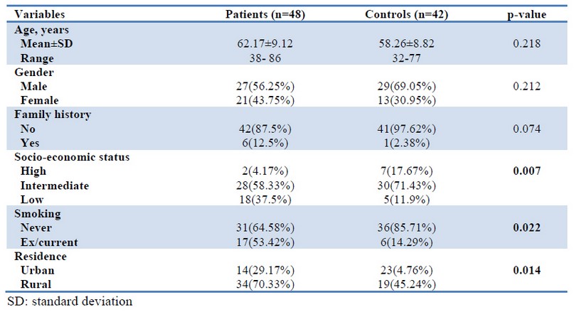
The mean age of the patients was 62.17±9.12 years, comparable to that of the controls (58.26±8.82 years) with no significant difference. Likewise, there were no significant differences between the two groups regarding gender distribution and family history of pTB. However, low SES, ex/current smokers, and rural residents were more frequent among patients (37.5%, 53.42% and 70.33%, respectively) than among controls (11.9%, 14.29% and 45.24%, respectively) with significant differences (Table 1).

Table 1. General characteristics of the study population.
Molecular Assay
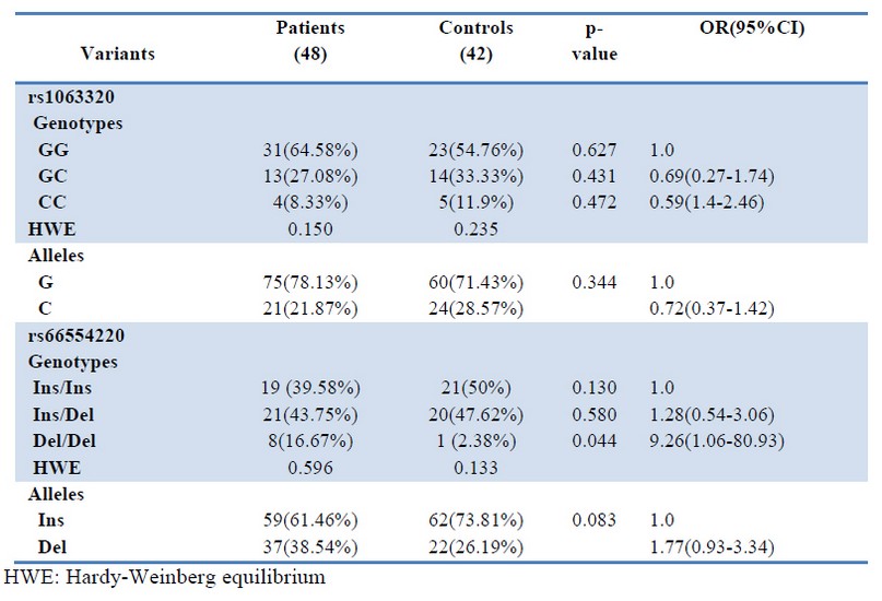
Each of rs1063320 (+3142G>C) and rs66554220 (14-bp Ins/Del) SNPs appeared in three genotypes. For rs1063320 (+3142G>C), these genotypes were GG, GC and CC, while the genotypes for rs66554220 (14-bp Ins/Del) were Ins/Ins, Ins/Del and Del/Del.
The frequency of different genotypes and alleles of both SNPs in either patients or controls followed the Hardy-Weinberg equilibrium. For rs1063320 (+3142G>C), patients and controls had a comparable rate of genotypes and alleles with no significant differences. On the other hand, the frequency of Del/Del genotype in patients (8.33%) was higher than that of controls (2.38%) with a significant difference (crude OR= 9.26, 95%CI=1.06-80.93, p=0.044). Although the del allele was remarkably more frequent in patients than controls (38.54% vs. 26.19%), the difference was not significant (crude OR= 1.77, 95%CI=0.93-3.34, p=0.083) (Table 2).

Table 2. The frequency of genotypes alleles of rs1063320 (+3142G>C) and rs66554220 (14-bp Ins/Del) in PTB patients and controls
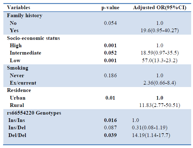
All factors that had a p-value<0.1according to the univariate analysis, were entered into the multivariate model. After adjusting for confounding factors, including smoking, family history, residence and SES, the multivariate analysis revealed a significant independent association between Del/Del genotype of rs1063320 (+3142G>C) SNP with the incidence of pTB (adjusted OR= 14.19, 95%CI=1.14-17.7, p=0.039). Furthermore, each of low SES and rural residence were also independent risk factors for pTB (adjusted OR= 57.0, 95%CI=13.3-23.2, p= 0.001 and adjusted OR= 11.83, 95%CI=2.77-50.51, p= 0.01) as shown in table 3.

Table 3. Multivariate analysis adjusted for confounding factors
Serum Level of Soluble HLA-G
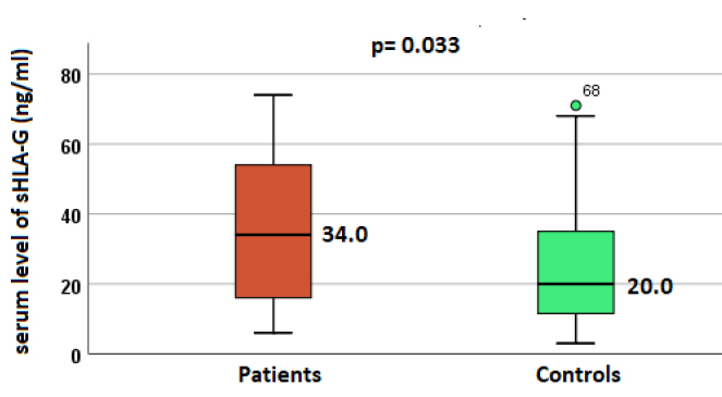
Median serum level of sHLA-G in pTB patients was 34.0 ng/ml (range 6.18-74.25 ng/ml), which was higher than that of controls (median 20 ng/ml, range 312-71.98 ng/ml) with a significant difference (Figure 1).

Figure 1. Median serum level of sHLA-G in patients and controls
DISCUSSION
In the present study, low SES, smoking and rural residence were significantly associated with susceptibility to pTB in univariate analysis. However, in multivariate analysis, smoking was not significant. The association of poverty with adverse health conditions was previously proved not only with TB but almost with all infectious diseases. Low SES is always associated with many predisposing factors for TB, such as low education level, high crowding, and low-quality water supply, which increase the chance for disease occurrence and transmission. Rural residents may have less access to health centers and low hygienic measures compared with urban residents. Thus, rural residents are more prone to infectious diseases, including TB, which can be a source of infections for other people because, in many cases, the patient is not got the proper treatment.
The most exciting finding in the present study was that the Del/Del genotype of the rs66554220 polymorphism was significantly associated with an increased risk of pTB (14.19,95%CI=1.14-17.7). This implies that individuals carrying this genotype have a 14-time greater risk of developing pTB than those carrying Ins/Ins genotype under the same circumstances. Most available studies addressing this polymorphism focused on non-infectious diseases. For example, García-González21 evaluated the 14-bp Del/Ins HLA-G polymorphism in acute coronary syndrome and type 2 diabetes mellitus (T2DM). The Ins/Ins genotype was associated with high blood pressure in diabetic patients. Another study found a significant association between the Ins/Ins genotype and coronary artery disease (CAD)22. The 14-bp insertion polymorphism was found to raise HLA-G mRNA instability, with a subsequent downregulation of HLA-G expression and reduction in membrane-bound and soluble mRNA isoforms production23. Svendsen et al. 24demonstrated that an increase in the expression of membrane-bound HLA-G1 coincided with a reduction of NK cytotoxicity in 14-bp/Ins transfected K562 cells compared to 14-bp/Del transfected cells. Furthermore, the study also showed that the HLA-G1 14-bp/Del allele was associated with a higher sHLA-G1/mHLA-G1 ratio than the 14-bp/Ins allele. Based on these observations, it can be speculated that the 14-bp/Ins associated with the protective role against pTB in the current study could be in charge of the main expression of membrane-bound isoforms of HLA-G, with eventual T-helper type 2 immune response and anti-inflammatory cytokines production.
Another critical finding in the present study was that sHLA-G was significantly higher in patients than in controls. Again most previous studies investigated this protein in non-infectious diseases. Fainardi et al.25investigated the variations in serum sHLA-I levels in multiple sclerosis (MS) patients under interferon (IFN) therapy. The authors demonstrated an elevation in serum sHLA-I levels one-month post-therapy. Furthermore, serum sHLA-I levels surged in responders compared with non-responder patients during the first trimester of treatment, suggesting that the favorable effect of interferon could be intermediated by increment in serum sHLA-I levels during the treatment course. This implies the immune suppression nature of this protein, which is incredibly beneficial in MS but harmful in TB. Many other studies supported this assumption that reported increased levels of HLA-G in breast cancer patients17,18,26. HLA-G protein is thought to have an immuno-inhibitory consequence on antigen-presenting cells (APCs), NK cells and T cells through the interactions of their specific inhibitory receptors, with KIR2DL4, which are expressed by almost all subsets of T-cells27. Additionally, as some anti-proinflammatory cytokines like interleukin (IL)-10 can boost HLA-G expression, which increases IL-10 production, it has been assumed that HLA-G antigens may have a role in immune modulation via promoting the immune switching from Th1 to Th2 response28.
CONCLUSIONS
Collectively, these data suggest that the del/del genotype of the rs66554220 polymorphism can be a risk factor for pTB by increasing HLA-G expression.
Conflict of interest
The authors declare that they have no conflict of interest
Acknowledgment
The authors are very grateful to all consultant clinic staff for respiratory diseases in Hilla/Iraq for their help in sample collection.
REFERENCES
1. Ogarkov, O.B.; Sinkov, V.V.; Medvedeva, T.V.; Gutnikova, M.Y.; Nekipelov, O.M.; Raevskaya, L.Y.; et al. polymorphism of genes of the renin-angiotensin system ACE, AT1R, and AT2R in patients with pulmonary tuberculosis. Mole Gene Microbiol Virol 2008;23(2):63-70
2. WHO 2010. Multidrug and extensively drug-resistant TB (M/XDR-TB): 2010 global report on surveillance and response. WHO/HTM/TB/2010.3 WHO Press, Geneva, Switzerland.
3. Möller, M.; Hoal, E.G. Current findings, challenges and novel approaches in human genetic susceptibility to tuberculosis. Tuberculosis (Edinb) 2010;90(2):71-83.
4. Mahasirimongkol, S.; Yanai, H.; Nishida, N.; Ridruechai, C.; Matsushita, I.; Ohashi, J. et al. Genome-wide SNP-based linkage analysis of tuberculosis in Thais. Genes Immun. 2009;10(1):77-83.
5. Png, E.; Alisjahbana, B.; Sahiratmadja, E.; Marzuki, S.; Nelwan, R.; Balabanova, Y. et al. A genome wide association study of pulmonary tuberculosis susceptibility in Indonesians. BMC Med Genet. 2012 Jan 13; 13(5): 10.
6. Dorling, A.; Monk, N.J.; Lechler, R.I. HLA-G inhibits the transendothelial migration of human NK cells. Eur J Immunol 2000;30(2):586–593
7. Donadi, EA.; Castelli, E.C.; Arnaiz-Villena, A.; Roger, M.; Rey, D.; Moreau, P. Implications of the Polymorphism of HLA-G on its function, regulation, evolution and disease association. Cellular Mole Life Sci 2011; 68(3):369–395
8. Lynge Nilsson, L.; Djurisic, S.; Hviid ,T.V. Controlling the immunological crosstalk during conception and pregnancy: HLA-G in reproduction. Front Immunol 2014;5:198.
9. Martelli-Palomino, G.; Pancotto, J.A.; Muniz, Y.C.; Mendes-Junior, C.T.; Castelli, E.C.; Massaro, J.D. et al. Polymorphic sites at the 3' untranslated region of the HLA-G gene are associated with differential HLA-G soluble levels in the Brazilian and French population. PLoS ONE. 2013;8:e71742.
10. Rousseau, P.; Le, Discorde, M.; Mouillot, G.; Marcou, C.; Carosella, E.D.; Moreau, P. The 14bp deletion-insertion polymorphism in the 3' UT region of the HLA-G gene influences HLA-G mRNA stability. Hum Immunol 2003;64(11):1005-1010
11. Chen, X.Y.; Yan, W.H.; Lin, A.; Xu, H.H.; Zhang, J.G.; Wang, X.X. The 14bp deletion polymorphisms in HLA-G gene play an important role in the expression of soluble HLA-G in plasma. Tissue Antigens. 2008;72:335–341.
12. Castelli, E.C.; Ramalho, J.; Porto, I.O.; Lima, T.H.; Felício, L.P.; Sabbagh, A. et al. Insights into HLA-G genetics provided by worldwide haplotype diversity. Front Immunol 2014;5:476.
13. Castelli, E.C.; Veiga-Castelli, L.C.; Yaghi, L.; Moreau, P.; Donadi, E.A. Transcriptional and posttranscriptional regulations of the HLA-G gene. J Immunol Res 2014;2014:734068.
14. Gautam, S.; Kumar, U.; Kumar, M.; Kanga, U.; Dada, R. Association of HLA-G 3'UTR Polymorphisms with Soluble HLA-G Levels and Disease Activity in Patients with Rheumatoid Arthritis: A Case-Control Study. Immunol Invest. 2020;49(1-2):88-105.
15. Zhou, S.; Liu, M.; Xia, Y.; Zhang, L.; Shao, L.; Wang, N.; Yu, W.; Ding, N.; Zhang, K,.;Liang, X. Association of the 3' untranslated region polymorphisms of HLA-G with susceptibility to chronic hepatitis C virus infection in the Chinese population. Hum Immunol. 2021 Sep 20:S0198-8859(21)00213-5.
16. Farghaly, W.M.; Saad Eldien, H.M.; Sayed, M.A. et al. The relationship of HLA-G 14-bp insertion/deletion genetic polymorphism to the risk of multiple sclerosis and its clinical phenotypes. Egypt J Neurol Psychiatry Neurosurg 2012;57:58.
17. Adler, H.; Sraub, C.; Frei, R. Comparison of BacT/ALERT 3D,Lowenstein-Jensen medium and Middlebrook 7H10/7H11 biplate for recovering mycobacteria from clinical specimens. Eur Clin Microbiol. Infect Dis, 2005;24(7):499-500.
18. Eskandari-Nasab, E.; Hashemi, M.; Hasani, S.S.; Omrani, M.; Taheri, M.; Mashhadi, M.A. Association between HLA-G 3'UTR 14-bp ins/del Polymorphism and susceptibility to breast cancer. Cancer Biomark 2013;13(4)253-259
19. Hashemi, M.; Sandoughi, M.; Fazeli, S.A., Bahari, G., Rezaei, M., Zakeri, Z. Evaluation of HLA-G 14bp ins/del and +3142G>C polymorphisms with susceptibility and early disease activity in rheumatoid arthritis. Adv Med 2016;2016:4985745.
20. Alsan, MM; Westerhaus, M.; Herce, M.; Nakashima, K.; Farmer, P.E. Poverty, global health, and infectious disease: lessons from Haiti and Rwanda. Infect Dis Clin North Am. 2011;25(3):611-ix.
21. Garcia-Gonzalez, I.J.; Valle, Y.; Rivas, F.; Figuera-Villanueva, L.E.; Munoz-Valle, J.F. Flores-Salinas HF, et al. The 14bp Del/Ins HLA-G polymorphism is related with high blood pressure in acute coronary syndrome and type 2 diabetes mellitus. Biomed Res Int 2014;2014:ID 898159
22. Boiocchi, C.; Bozzini, S.; Zorzetto. M.; Pelissero, G.; Cuccia, M.; Falcone, C. Association between two polymorphisms in the HLA-G gene and angiographic coronary artery disease. Mole Med Rep 2012;5(5):1141–1145.
23. Hviid, T.V.F. HLA-G in human reproduction: aspects of genetics, function and pregnancy complications. Hum Reprod Update. 2006; 12:209–232.
24. Svendsen, S.G.; Hantash, B.M.; Zhao, L.; Faber, C.; Bzorek, M.; Nissen, M.H. et al. The expression and functional activity of membrane-bound human leukocyte antigen-G1 are influenced by the 3’-untranslated region. Hum Immunol. 2013; 74:818–827.
25. Fainardi, E.; Rizzo, R.; Melchiorri, L.; Castellazzi, M.; Govoni, V.; Caniatti, L. et al. Beneficial effect of interferon-1β treatment in patients with relapsing-remitting multiple sclerosis is associated with an increase in serum levels of soluble HLA-I molecules during the first 3 months of therapy. J Neuroimmunol 2004;148:206–11.
26. Zidi, I.; Rizzo, R.; Bouaziz, A.; Laaribi, A.B.; Zidi, N.; Di, Luca, D. et al. sHLA-G1 and HLA-G5 levels are decreased in Tunisian women with multiple abortion. Hum Immunol. 2016;77: 342–345.
27. Carosella, E.D.; Favier, B.; Rouas-Freiss, N.; Moreau. P.; LeMaoult, J. Beyond the increasing complexity of the immunomodulatory HLA-G molecules. Blood 2008;111:4862–4670.
28. Carosella, E.D.; Moreau, P.; Aractingi, S.; Rouas-Freiss, N. HLA-G: a shield against inflammatory aggression. Trends Immunol 2001;22:553–555.
Received: 2 March 2022 / Accepted: 31 April 2022 / Published:15 Agoust 2022
Citation: Bushra J. Al –Tamimi, Qasim S. Al-Mayah, Hadeel S. Abd-Alwahab. Study of HLA-G gene polymorphism and serum level of soluble HLA-G in patients infected with Mycobacterium tuberculosis. Revis Bionatura 2022;7(3) 24. http://dx.doi.org/10.21931/RB/2022.07.03.23
