2022.07.03.39
Files > Volume 7 > Vol 7 No 3 2022
Detection of morphometrical, histological and histochemical characteristics of lung and trachea in adult local squirrel (Sciurus anomalus)

Iman Mousa Khaleel 1* ,Khalid Ibrahim Abd Alkhazraji2 , Mahdi Abdul kreem Atiea 3
1,3Department of Anatomy and Histology, College of Veterinary Medicine, University of Baghdad, Iraq
2 Department of Anatomy and Histology, College of Veterinary Medicine, University of Diyala, Iraq
Corresponding author: [email protected], https://orcid.org/0000-0001-6038- 1608
Available from: http://dx.doi.org/10.21931/RB/2022.07.03.39
ABSTRACT
The present study aimed to identify the topography, morphology, histochemistry and histology of lung structures, bronchial divisions and trachea in adult local Squirrel (Sciurus anomalus) as a species inhabited in Iraqi environments. This work was conducted on thirty local Squirrel of both sexes (15) males and (15) females were divided into three equal groups, first for http://wsx5customurl.comanatomical perceptions, second to resin cast technique and the third for histological study. Anatomically, in both sexes, the trachea appeared as a cartilaginous structure consisting of flexible cartilaginous rings, C-like, connected by annular ligaments. It begins at the end of the cricoid cartilage from the level of the second cervical vertebra to the fourth thoracic vertebral plane; eventually, it splits into the right and left main bronchi. Count tracheal rings, the entire weight of the lung, full length, the diameter of the trachea and right and left bronchi. The trachea in females was slightly less than in males but not significantly. The bronchial tree was detected in resin cast, which was constructed of the trachea divided into left and right primary bronchi (Main bronchi), the right one was split into four secondary bronchi to enter the right lobes and two secondary bronchi to the left one. The left lung contains one lobe, whereas four lobes were observed in the right. Histologically, the wall of the trachea consists of four layers. Epithelial cells of ciliated pseudostratified columnar and goblet cells that reacted positively with PAS were covered in the mucosa. Submucosa was devoid of the tracheal glands. The Trachealis muscle is connected from the outer aspect of rings.
Similarly to the trachea, the primary bronchi are structured but smaller in diameter; they break up within the lung into primary, secondary and tertiary bronchi, then it terminates in respiratory bronchioles that contain Clara cells and open at the end in the sacs of alveoli. Two types of pneumocystis were observed lining the alveoli. The current study concluded anatomically and histologically that there were no significant differences between males and females of local squirrels. The lobulation of the lung in squirrels is different from other animals. The trachea and lung histologically resembled numerous animals, however, the wall had micro morphometric changes. But, the surface lining cells of the tracheal and bronchi mucosa secrete neutral mucin, with no submucosal glands in the tracheal wall.
Keywords: Trachea, Local Squirrel, Bronchial Tree, Histochemical, Lung.
INTRODUCTION
Respiratory system a critical system that play a vital role in the ventilation system which limits the gases exchanging via the respiratory structure and outside air 1. Olfaction, Vocalization and regulation of the body temperature regard another respiratory activation besides the ventilation, consisting of gas exchange and conduction 2. The respiratory system comprises many anatomical structures divided into conducting and respiratory parts in mammals. The terminal site of this system is alveoli which are responsible for gas exchanging 3. The distribution of the air and blood in a vast space by an extensive network of airways and blood vessels will assist in gas exchanging 4. In most domestic animals, the trachea wall is made of (4) layers; Mucosa is lined by pseudostratified columnar epithelium, submucosa, muscularis and adventitia. The incomplete hyaline cartilaginous rings support these layers 5. The conducting and respiratory portions have different anatomical characteristics among species and individuals 6. The morphometrical studies of the trachea and lung are critical due to the vital role of the trachea during breathing and intubation processes through general anesthesia7. The Squirrel (Sciurus anomalus ) belongs to the order Rodentia, family; Sciuridae, Genus; Sciurus and Species; Sciurus anomalus. 8 indicate that this species is characterized by hibernation, which lasts from early autumn to spring, and the animal remains hidden underground. Significant alterations, whether in the immunological state or in many sides of lung mechanization, could occur to the lung of hibernating squirrels9,10. Many authors have studied the anatomical and histological lung structure in small and lab animals like mice, rats, and guinea pigs11,12,13,14. There is a lack of literature on the histological and morphological features of the lung and trachea in local Squirrels. Therefore we studied the morphological, morphometrical, histological, and histochemical features of the trachea and lungs and described the bronchial tree using the resin technique in this animal.
MATERIALS AND METHODS
Thirty healthy adult squirrels of both sexes were divided into three equal groups, each one containing ten animals (5 male and 5 female squirrel) with an average weight (214) g. The studied animals were obtained from local supplier in Al-gazil market. The first group was used for topography, gross anatomy of trachea and lung, length and diameter of trachea measured by electronic digital vernier caliber and ruler, weight by sensitive balance and counting of tracheal ring numbers. In contrast, the second group was used for the bronchial tree by making corrosive resin cast, and the third group was used for the histochemical and histological examination.
Corrosive cast technique
The resin injection was done by hand pressure through a syringe of (20) ml; the specimens were injected with ( 2 ) ml of resin. After complete solidification, the specimens were macerated through a solution of (40%) KOH for (3-4) days, washed and dried by hot air until become ready for examination according to 15 .
Histological study
The specimen was taken from the trachea, primary left and right bronchus and lobes of both lungs, fixed in 10% buffered formalin, washed with water, and dehydrated in a series of alcohol (70%, 80%, 90%, 100%, 100% ) in that order, two-run for each, left in each concentration for an hour. Cleared in xylene, infiltrated by molten paraffin-embedded in paraffin wax slandered procedures. I was sectioned in (6-7) µm. Stained by Harris Hematoxylin and Eosin stain and Periodic Acid Schiff, Alcian blue (PH 2.5) according to 16 .
Statistical Analysis
The statistical analysis was applied using two ways (ANOVA) and means a significant difference at (P≤0.05). In using a statistical package for social sciences (T-Test).
RESULTS
The trachea of squirrels in both sexes looked like a flexible, hollow cylindrical tube made of numerous rings of hyaline cartilage that was confirmed by an annular ligament, their ring edges and muscle filled it, according to this study (Fig. 1). The trachea was elastic structure lie on the ventral side of the neck at midline. It extends between cricoid cartilage of the larynx to the (carina) at the fourth thoracic vertebra. Then it was bifurcate at the level of the 4th thoracic vertebra into the right and left principal bronchi (Fig.2).
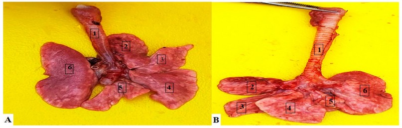
Figure.1. Gross anatomy of trachea and lung (A: dorsal view, B: ventral view) in female Squirrel Shows:
1-trachea.2-right apical lobe.3-right middle lobe.4- right caudal lobe.5-left accessory lobe.4-left lung
Table 1 summarizes the cartilaginous rings that appeared as C-shape structures connected by tracheal muscle at the two ends. The mean tracheal length in males was higher than that of females, and the mean number of tracheal rings was less than in females. The tracheal rings were rounded in shape or semi-circular in their cross-section. The length, and ring numbers in the cervical and thoracic region of the trachea, in males were higher than of females but not significantly. The mean tracheal and right principal bronchus diameters in males were higher than that of females, whereas the left principal bronchus diameter in females was higher than that in males. Significantly no difference between them at (P≤0. 05) in all anatomical parameters. Topographically, the trachea was separated into cervical and thoracic parts (Fig. 2).
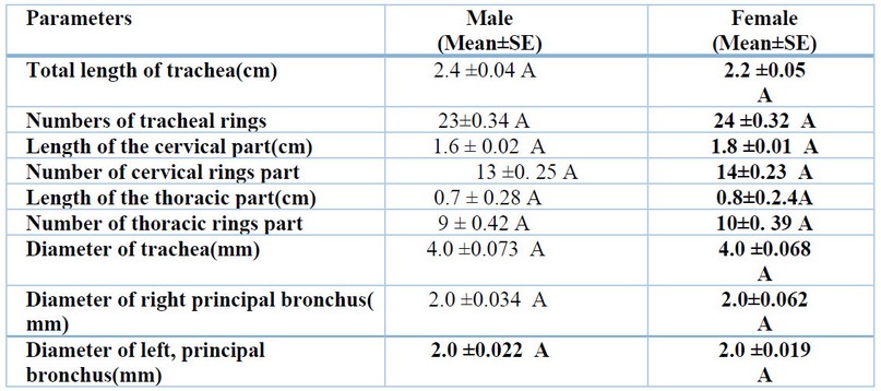
Table 1. Morphometric measurements of the trachea of Squirrel in both sexes (Mean±SE).
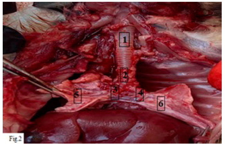
Figure 2. The gross anatomy of the male Squirrel trachea Shows 1-Cervical part, 2- Thoracic region, 3- 4 right& left bronchus, 5- Right lung. 6- Left lung
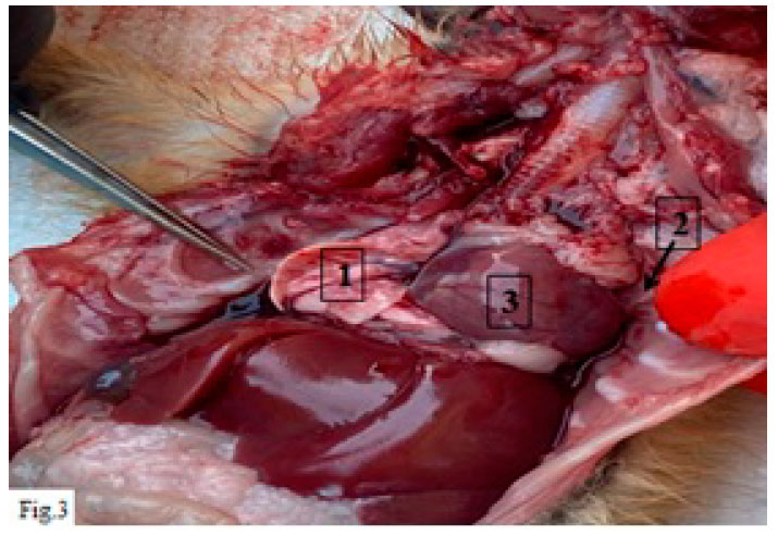
Figure 3. The gross anatomy of the male Squirrel trachea Shows 1- Right lung. 2- Left lung,3- Heart.
The Lung
As in domestic animals, the lung in both sexes appear as two reddish pink spongy structures surrounding the heart, and their shape corresponds to the thoracic cavity shape (Fig.3). In both sexes, the right and left lungs have an apex, base, dorsal, ventral and caudal borders. The ventral and caudal edges were flatted and pointed, whereas the dorsal border was thick and rounded. The right lung apex was large and round, whereas the left was smaller and pointed. Both lungs have three convex and narrow surfaces; costal, medial and diaphragmatic. Interlobar fissures separated the lung into distinct lobes. The right lung contains apical, middle, caudal, as well as accessory lobes, which are partially divided into two small lobes (Fig. 3). Left lung is made up of one lobe only (Fig. 1). Currently, the lungs of both sexes do not have clear sub-pleural pulmonary lobules. The mean body weight, total lung weight, and right and left lung weights in males and females were listed in (Table 2). These measurements were higher in males than females but not significant at P≤0.05 (Table 2). There is no sign in the weights of the right and left lungs in males compared with females.
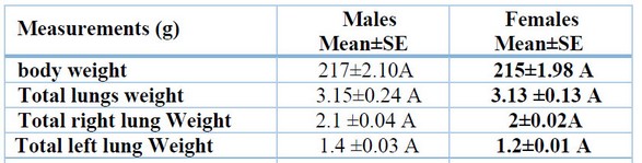
Table 2. Morphometric measurements (g) of the lung in both sexes of Squirrel (Mean±SE)
The corrosive cast technique displays the trachea and bronchial tree form that the trachea consists of left and right (principal or main) bronchi. The left spilled to the left apical bronchus and caudal bronchus for the left apical and caudal ends of the left lobe (Fig. 4). The other main bronchi, right, observed to split into 4 bronchi as an accessory, caudal, middle and apical (Fig. 4). From the right primary bronchus, the first branch enter the apical lobe. Both central and accessory bronchus arise at the same level beyond the apical bronchus and join the middle and additional lobes, respectively. In contrast, the caudal lobe receives bronchi of the right caudal (Fig. 4 ). The bronchiole of tertiary and secondary arises from the division of tertiary and secondary bronchus, which arises from the right main bronchus.
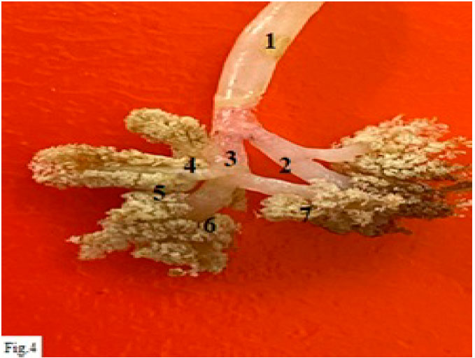
Figure 4. The corrosive cast technique for the bronchial tree Shows: 1-Trachea.2-right principal bronchus, 3-left principal bronchus.4- R. apical primary bronchi.5-R.Middle primary bronchi. 6-R.Caudal primary bronchi, 7-R.Accessory primary bronchi.
Histological Results
In both sexes, the trachea of Squirrel was conformable and appeared as a solid, hollow tubular structure consisting of the mucosa, submucosa, cartilaginous layer, and adventitia. The mucosa is lined by Pseudostratified ciliated columnar epithelial cells, which contain (ciliated columnar cells, goblet cells and basal cells) resting on the basement membrane. The main cell types were the ciliated columnar cells found as tall columnar cells with apical cilia. These cells were slightly stained cytoplasm and apical large oval nuclei (Fig. 5). Numerous goblet cells have global shapes and basally located nuclei. It gives a positive reaction towards PAS, giving a magenta color (Fig.6), due to its secrete a mucopolysaccharide substance. The mucous secretion may act as an epithelial protective barrier. The basal cells display small triangular cells resting on the basement membrane but don't reach the lumen. A thin, loose connective tissue layer made from collagenous and elastic fibers represents lamina propria. Muscular mucosa is observed as the very thin layer of smooth muscle fibers.
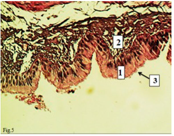
Figure 5. Histological section of the trachea in male Squirrel Shows Cilia (black arrow) 1-epithelium 3-Lamina propria H and E stain X40
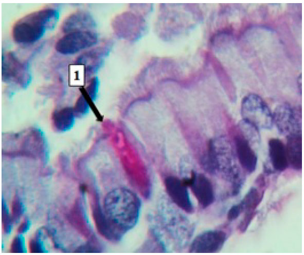
Figure 6. Histological section of the trachea in female Squirrel Shows:1- Goblet cells PAS stain X100
Loose connective tissue represents tunica submucosa devoid of submucosal glands (Fig. 7). The muscular cartilaginous layer merges with hyaline cartilage perichondrium and fibroblastic tissue that is observed between the cartilaginous rings. The chondrocytes present in an amorphous matrix in hyaline cartilage (Fig.7),
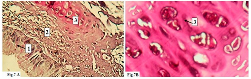
Figure 7. Histological section of the trachea in female Squirrel Show.1-Mucosa 2- Submucosa. 3-Hyalin cartilage. Submucosal glands absent. H and E stain ( A:X 100, B: X400)
The cartilaginous rings were C-shaped, opened dorsally, and filled with connective tissue and a smooth tracheal muscle connecting the rings' outer side (Fig. 8). Tunica adventitia coated the cartilage and displayed a loose connective tissue. The tracheal bifurcation occurs at the carina forming the right and left, principal bronchi which histologically formed from; mucosa which covered with the epithelium of pseudostratified ciliated columnar, submucosa represented via loose connective tissue devoid of glands. The fibro-cartilaginous layer contains hyaline cartilage, and the adventitia is composed of loose connective tissue. The primary bronchi lead to the secondary bronchi and tertiary bronchi. The secondary bronchi were made up of folded mucosa coated by ciliated pseudostratified columnar epithelial cells with goblet cells which react positively towards the PAS stain. Thin lamina propria, bundles of smooth muscle fibers split up the lamina propria from the submucosa. The bronchial glands were absent. The tracheal and primary bronchi cartilaginous rings were replaced by separated hyaline cartilage plates in the secondary bronchi. Adventitia contains small blood vessels and is surrounded immediately by the lung parenchyma. The secondary bronchi lead to tertiary bronchi that are distinguished by the folded mucosa lined by simple columnar ciliated epithelial with goblet cells which react positively for the PAS procedure. The epithelium of smaller bronchi was low simple ciliated columnar epithelium. Lamina propria is made from elastic fibers, smooth muscle and connective tissue. Irregular hyaline cartilage plates smaller than those of the secondary bronchi (Fig. 9).
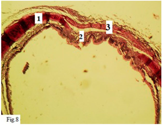
Figure 8. The histological section in the trachea of a male Squirrel Shows 1-Hyaline cartilage. 2-Tunica mucosa3Trachealis muscle. H and E Stain(X4).
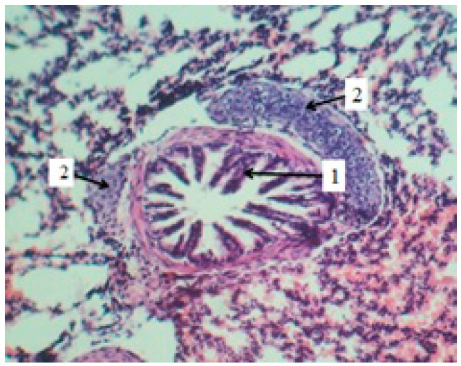
Figure 9. Histological section of the lung in female Squirrel Shows Tertiary bronchiole:1- Folded mucosa, 2- plates of hyaline cartilage PAS stain (X 40).
The submucosal connective tissue was merged with the adventitia. The lining epithelium of the bronchioles was changed to become low simple columnar ciliated epithelial cells and goblet cells. Clara cells appeared as a dome shape containing light cytoplasm with a central nucleus (Fig.10). Lamina propria showed as thin, loose connective tissue enclosed by a layer of smooth muscle fibers. The cartilaginous plates were absent (Fig.10). Lining mucosa epithelium in the large terminal bronchioles was pseudostratified columnar ciliated epithelial cells but became simple ciliated columnar to simple cuboidal cells in the smaller terminal bronchioles, and the goblet cells were devoid (Fig. 11).
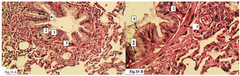
Figure 10. Histological section of the lung in male Squirrel shows bronchiole:1-simple columnar ciliated epithelium, 2-Clara cells.3-Smooth muscle Fibers, 4- goblet cell PAS Stain A-X40, B-X100.
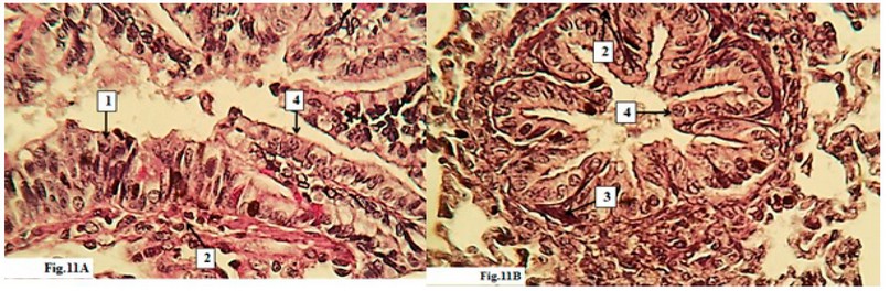
Figure 11. Histological section of the lung in female Squirrel shows: Large terminal bronchiole shows: A- 1-pseudostratified prismatic ciliated epithelial cells, 2- Lamina propria, 3- Smooth muscle fibers, B- Simple 4-cuboidal cells, PAS Stain X400, B-Hand E Stain X 100.
Lamina propria lack smooth muscle fibers. The terminal bronchioles open into the respiratory bronchioles, which line by a simple cuboidal epithelium except in the spaces of alveoli (out pocketing) on their walls. The respiratory bronchioles were opened directly into alveolar ducts that opened into the alveolar sacs and the alveoli (Fig.12). Alveolar ducts were long, straight, forward tubular structures, line by simple squamous epithelial cells with many out pocketing of alveoli. Alveolar ducts lead to the alveolar sacs, which consists of alveolar cluster around collective air space (Fig. 12),
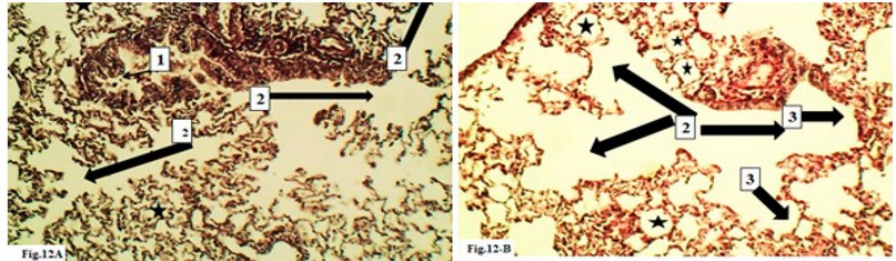
Figure 12. Histological section of the lung in female Squirrel shows 1- Respiratory bronchiole, 2-alveolar duct, 3- alveolar sac, alveoli (black star) A-H and E X40, B-PAS X40.
The lung parenchyma of Squirrel was made of alveoli, interalveolar septa and blood capillaries; the alveoli appeared as small spherical air spaces lined by very thin epithelium consisting mainly of type I pneumocyte (simple squamous cells) with centrally situated nuclei. Another type of cell showed cuboidal to rounded scattered amongst the first kind named pneumocyte type II (Fig.13). The interalveolar septa was constructed of a single layer of epithelium and a skinny subepithelial layer of connective tissue fibers and blood capillaries. The visceral pleura was made of a thin connective tissue covered by mesothelium (Fig.14). The devastation of alveolar walls as local damage were characteristic features for parenchyma in the lung of studied squirrels.
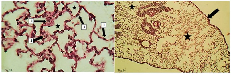
Figure 13. Histological section of the lung in male Squirrel Shows 1- TypeI Pneumocyte (red arrows), Type II Pneumocyt, Alveoli (black star )PAS Stain X100.
Figure 14. Histological section of the lung in female Squirrel Shows Visceral Plura (black arrow), H and E stain X40.
DISCUSSION
The gross anatomy of the trachea in both sexes of squirrels showed as a flexible, hollow cylindrical tube consisting of many hyaline cartilage rings confirmed by an annular ligament and their ring edges filled by smooth muscle. Similar to that reported in local cats by17. The location and extension of the trachea between the cricoid cartilage to the carina and its were agreed with the result reported in cats (Files catus) by 18. Its bifurcation into the right and principal bronchi at the level of 4th thoracic vertebra was agreed with that observed in guinea pigs (Cavia porcella ) by 14. The C-shape structure of the cartilaginous rings connected by the tracheal muscle at the two ends was similar to that observed by 19, but disagree with that reported in red sokoto goat (U) shaped structure 20. The tracheal lengths and ring numbers varied between males and females but not significantly. The tracheal measurements and ring numbers vary within species and among species as in camel( Camelus dromedaeies), the tracheal length was 92-101cm by 21, monkey squirrel was (30.25-32.8) ring22.
The general shape, color and relationship of the lung in both sexes was similar to the finding by 13 in mole rats and by 23 in gazelle. The gross anatomy of the right and left lungs, as found in rabbits by24 and small ruminants 25. Interlobar fissures separated the lung into distinct lobes; the number of lobes in the right and left lobes in both sexes of Squirrel was agreed with26 in mice but disagreed with that reported in rabbits in which the left lung was composed of two lobes; anterior and posterior lobes 27. The lungs of both sexes do not have clear sub-pleural pulmonary lobules that differ from those of small ruminants 25 but agree with that found in rabbits 28. The corrosive cast technique displayed the form of the trachea and bronchial tree, which differed from that observed by 19 Iraqi weasels (Herpestes javanicus), in which the left primary bronchi were divided into apical, middle and caudal bronchi to the apical, middle and caudal lobes. The other main bronchi, right, was split into four bronchi as an accessory, caudal, middle and apical, similar to that observed by 29 in (Cryptosis parva). The branches of the right primary bronchus were agreed with that found in rabbits by24. The bronchiole of tertiary and secondary arises from the division of tertiary and secondary bronchus, which rises from the right main bronchus. These differences may be due to species differences and environmental adaptation.
Histological discussion
In both sexes of Squirrel, the trachea was conformable and appeared as a solid, hollow tubular structure consisting of mucosa, submucosa and cartilaginous layer and adventitia, as reported in many domestic animals 30. The lining epithelium of the mucosa was Pseudostratified ciliated columnar epithelial cells, including the goblet cells, as observed in goats by 31. Numerous goblet cells have a global shape with basally located nuclei. It positively reacts toward PAS due to its secrete a mucopolysaccharide substance. The mucous secretion may act as an epithelial protective barrier. At the same time, the basal cells appeared as small triangular cells that rested on the basement membrane but didn't reach the lumen, similar to that reported in goats by 32. The lamina propria and muscular mucosa structure were observed to be identical to that reported in the Iraqi weasel (Herpestes javanicus) by19. Whereas the submucosa was represented by loose connective tissue devoid of submucosal glands, this finding disagreed with that reported in equines in which the submucosal mucous glands were very few 33, but differed from that observed by 34 in sheep in which the submucosal glands were mixed. The mucus has an essential role in preventing dust accumulation 35. The submucosal gland contains goblet cells that secrete mucus which moisture and lubricate and provides an average mucociliary clearance and acts as protection to the trachea 36.
The muscular cartilaginous layer merges with hyaline cartilage perichondrium and fibroblastic tissue which are present between the cartilaginous rings, as reported in gazelle by 23. The characteristic of the cartilaginous rings was agreed with that found in other species, as in Arabian Oryx7. The tunica adventitia covered the cartilage and displayed a loose connective tissue; a similar result was documented by 5. The fibrocartilaginous layer contains hyaline cartilage, and the adventitia is composed of loose connective tissue; the same finding was recorded in the ferret in red fox by 37 who reported that there were tracheal or bronchial glands. The tracheal and primary bronchi cartilaginous rings were replaced by separated hyaline cartilage plates in the secondary bronchi. Adventitia contains tiny blood vessels and is surrounded immediately by the lung parenchyma; this finding was reported in other domesticated animals as 23 in gazelle. The secondary bronchi lead to tertiary bronchi with the same histological structure as found in the rat by 13. At the same time, the lining epithelium and lamina propria of the bronchioles were similar to those recorded in rhesus by 38. The lining epithelium of mucosa in the large terminal bronchioles was a pseudostratified columnar ciliated epithelial cells, then became simple ciliated columnar to simple cuboidal cells in the smaller terminal bronchioles, 39 reported that the terminal bronchioles were lined by simple cuboidal epithelium. The terminal bronchioles opened into the respiratory bronchioles, which lined by a simple cuboidal epithelium except in the spaces of alveoli on their walls; this was identical to that observed in Sambar deer by 40. The respiratory bronchioles were opened directly into alveolar ducts that opened into the alveolar sacs. The alveoli had similar results reported in other animals, such as in a nocturnal burrowing rodent 41. The microscopic structure of alveolar ducts and alveolar sacs was identical to that in guinea pigs by 14. The lung parenchyma in Squirrel, constructed of alveoli, inter alveolar septa and blood capillaries, and the alveoli lined by pneumocyte type I and peumocyte type II were previously observed in rats by 13. The interalveolar septa were constructed of a single layer of epithelium and a skinny sub-epithelial layer of connective tissue fibers and blood capillaries; a similar result was reported in goat by2. The devastation of alveolar walls as local damage were characteristic features for parenchyma in the lung of studied squirrels as observed in the rat by 13.
CONCLUSION
The current study concluded anatomically, and histologically there were no significant differences between male and female local squirrels. The trachea appeared as a cartilaginous structure consisting of flexible cartilaginous rings in C- like. The bronchial tree is constructed of the trachea, divided into left and right primary bronchi; the right one was split into four secondary bronchi to enter the right lobes, while two secondary bronchi to the left on. In Squirrel, the lobulation of the lung was different from that observed in other mammals. Histologically, the trachea and lung was similar to that reported in many mammals, but with some micro morphometrical differences in its wall. The histochemical findings show that the goblet cells in the tracheal and bronchi mucosa surface lining cells secret neutral mucin, and there are no submucosal glands in the tracheal wall.
Author conflict: No conflict
Acknowledgment: Thanks going for all who support us.
Conflict between authors: No conflict
Funds: self by authors
REFERENCES
1. Maton A, Hopkins JS, Johnson CW, McLaughlin MQ, Warner D, Lahart WJ. Human Biology and Health. Englewood Cliffs: Prentice Hall. 2010 : 108-118.
2. Baba MA, Choudhary AR. Histomorphology of the pulmonary alveoli of goat (Capra hircus). Veterinary World. 2008;1(10):312.
3. Maina JN. The morphology and morphometry of the adult normal baboon lung (Papio anubis). Journal of anatomy. 1987;150:229.
4. Yousif NH, Dawood MS. Morphometric Comparative Anatomical Study Of Lower Respiratory Tract Between Sheep (Ovis aris) And Goat (Caprus hircus) in Baghdad provence. Kufa Journal For Veterinary Medical Sciences. 2019;10(2).
5. Steiner M. The physiological basis of respiratory disease: Hamid, Shannon, Martin: Published by BC Decker: ISBN: 1550092367 Price:£ 58.99 (Hardcover). Chronic Respiratory Disease. 2008;5(2):121-.
6. Chunder R, Nandi S, Guha R, Satyanarayana N. A morphometric study of human trachea and principal bronchi in different age groups in both sexes and its clinical implications. Nepal Med Coll J. 2010;12(4):207-14.
7. Al-Zhgoul MB, Dalab AH, Abdulhakeem E, Ismail ZB, Thanain AT, AL-ZHGOUL MB, DALAB A, ABDULHAKEEM E, ISMAIL Z, THANAIN A. Arabian oryx (Oryx leucoryx) trachea: a descriptive and morphometric analysis. Int. J. Morphol. 2013;31(3):813-8.
8. Millesi E, Huber S, Dittami J, Hoffmann I, Daan S. Parameters of mating effort and success in male European ground squirrels, Spermophilus citellus. Ethology. 1998;104(4):298-313.
9. Milsom WK, Reid WD. Pulmonary mechanics of hibernating squirrels (Spermophilus lateralis). Respiration physiology. 1995;101(3):311-20.
10. Bohr M, Brooks AR, Kurtz CC. Hibernation induces immune changes in the lung of 13-lined ground squirrels (Ictidomys tridecemlineatus). Developmental & Comparative Immunology. 2014;47(2):178-84.
11. Zhang L, Li D, Luo S. Non-invasive microstructure and morphology investigation of the mouse lung: qualitative description and quantitative measurement. PLoS One. 2011;6(2):e17400.
12. Ilgun R, Yoldas A, Kuru N, Özkan ZE. Macroscopic anatomy of the lower respiratory system in mole rats (Spalax leucodon). Anatomia, histologia, embryologia. 2014;43(6):474-81.
13 Koptev M, Pronina O, Danilchenko S, Avetikov D, Stavitskiy S. Histological features of rats' normal lung tissue.
14. Mutlak BH, Al-Jawadi AA, Mnati IM. Morphological study of bronchial tree and lung in Iraqi weasel (Herpestes javanicus). InJournal of Physics: Conference Series 2021 (Vol. 1879, No. 2, p. 022044). IOP Publishing.
15. Abd Alameer SM. Anatomical Study of the Tracheal Cast and Lung in Local Breed Cats Felis Cactus Domesticus. L. Basrah Journal of Veterinary Research. 2013;12(2).
16. Bancroft JD, Gamble M, editors. Theory and practice of histological techniques. Elsevier health sciences; 2008.
17. Nasser RA, Mirhish SM. Histological Study Of Lower Respiratorsystem On Cats (Felis Catus Domesticus. L). In3rd Scientific Conference-College Of Veterinary Medicine-University Of Tikrit 2016.
18. Caccamo R, Twedt DC, Buracco P, McKiernan BC. Endoscopic bronchial anatomy in the cat. Journal of Feline medicine and surgery. 2007;9(2):140-9.
19. Mutlak BH, Al-Jawadi AA, Mnati IM. Morphological study of bronchial tree and lung in Iraqi weasel (Herpestes javanicus). InJournal of Physics: Conference Series 2021 (Vol. 1879, No. 2, p. 022044). IOP Publishing.
20. Nzalak JO, Ibe CS, Salami SO, Umosen AD, Ali M, Byanet O, Maidawa S, Imam J. Macroscopic Studies of the Lower Respiratory System of the Red Sokoto Goat (Capra aegagrus hircus). Journal of Veterinary Anatomy. 2013;6(1):47-52.
21. Hussein AJ, Zahra IA. Morphological, histological and histochemical study of trachea of one hump camel (Camelus dromedaries) in south of iraq. InFirst Iraqi Colloquium on camel Diseases and Management 2016 (Vol. 1, pp. 19-25).
22. Pinheiro LL, Lima AR, Muniz JA, Imbeloni A, Fioreto ET, Fontes RF, Cabral R, Branco É. Anatomy and morphometric aspects of the trachea of Saimiri sciureus Linnaeus, 1758: knowledge for emergency procedures. Anais da Academia Brasileira de Ciências. 2012;84:973-7.
23. Jumaah YR. Histomorphological Study of Trachea and Lung in adult indigenous Gazelle (Gazella subgutturosa) (Doctoral dissertation, council of Veterinary Medicine College, university of Baghdad).
24. Al-Anbaki AA. Anatomical and histological and radiological study of trachea and lungs in domestically rabbits (Doctoral dissertation, Vet. Med. Collage, University. of Baghdad).
25. Yousif NH, Dawood MS. Morphometric Comparative Anatomical Study Of Lower Respiratory Tract Between Sheep (Ovis aris) And Goat (Caprus hircus) in Baghdad provence. Kufa Journal For Veterinary Medical Sciences. 2019;10(2).
26. Thiesse J, Namati E, Sieren JC, Smith AR, Reinhardt JM, Hoffman EA, McLennan G. Lung structure phenotype variation in inbred mouse strains revealed through in vivo micro-CT imaging. Journal of applied physiology. 2010;109(6):1960-8.
27. Ramchandani R, Bates JH, Shen X, Suki B, Tepper RS. Airway branching morphology of mature and immature rabbit lungs. Journal of Applied Physiology. 2001;90(4):1584-92.
28. Autifi MA, El-Banna AK, Ebaid AE. Morphological study of rabbit lung, bronchial tree and pulmonary vessels using corrosion cast technique. Al-Azhar Assiut Med. J. 2015;13:41-51.
29. Arodaki F, Khamas W, Darmani N, Al‐Tikriti M. Histological characteristics of the tracheobronchial tree of the Least Shrew (Cryptotis parva). Anatomia, histologia, embryologia. 2017;46(4):405-9.
30. Uemura EE. Fundamentals of canine neuroanatomy and neurophysiology. John Wiley & Sons; 2015.
31 Habib RS, Mahammed FS. Histomorphological and radiological study of trachea and lungs of Angora goat (capra ibex) (Doctoral dissertation, M. Sc. thesis, Veterinary Medicine College, University of Duhok).
32. Kahwa CK, Purton M. Histological and histochemical study of epithelial lining of the respiratory tract in adult goats. Small ruminant research. 1996;20(2):181-6.
33. Widdicombe JH, Pecson IS. Distribution and numbers of mucous glands in the horse trachea. Equine veterinary journal. 2002;34(6):630-3.
34. Al-Umeri SK. Grossly and microscopic study of the trachea and bronchial tree in the local sheep (Ovis aris). Bas J Vet Res. 2015;14:145-55.
35. Thornton DJ, Sheehan JK. From mucins to mucus: toward a more coherent understanding of this essential barrier. Proceedings of the American Thoracic Society. 2004;1(1):54-61.
36. Moore KL, Dalley AF. Clinically oriented anatomy. Wolters kluwer india Pvt Ltd; 2018.
37. Moussa EA, Hassan SA. Histology and Scanning Electron Microscopy of the Lower Respiratory Tract in the Adult Red Fox (Vulpes vulpes). International Journal of Morphology. 2015 ;33(1).
38. Hyde DM, Hamid Q, Irvin CG. Anatomy, pathology, and physiology of the tracheobronchial tree: emphasis on the distal airways. Journal of Allergy and Clinical Immunology. 2009 ;124(6):S72-7.
39. Kalita A. Histomorphological study of the respiratory system of Mizo local pig (Zo Vawk). Asian Journal of Biomedical and Pharmaceutical Sciences. 2014;4(29):50.
40. Indu VR, Indu KL, Lucy KM, Harshan KR, Chungath JJ, Thankachen KC, Thankachen KC. Histomorpholoy of the pulmonary acinus in Sambar deer (Cervus Unicolor). Indian J Anim Res Indian J. Anim. Res. 2012;46(2):127-31.
41. Ibe CS, Salami SO, Onyeanusi BI. Macroscopic anatomy of the lower respiratory system in a nocturnal burrowing rodent: African giant pouched rat (Cricetomys gambianus, Waterhouse 1840). Anatomia, Histologia, Embryologia. 2011 Apr;40(2):112-9.
Received: 7 January 2022 / Accepted: 2 May 2022 / Published:15 August 2022
Citation: I M Khaleel, Abd Alkhazraji K I , kreem Atiea M A. Detection of morphometrical, histological and histochemical characteristics of lung and trachea in adult local squirrel (Sciurus anomalus). Revis Bionatura 2022;7(3) 39. http://dx.doi.org/10.21931/RB/2022.07.03.39