2023.08.02.57
Files > Volume 8 > Vol 8 No 2 2023
Phenotypic and Molecular Investigation of Streptococcus pneumoniae pneumolysin

Roaa A. Thamer 1,* , Amera M.Al-Rawi 2
1 Department of Biology / College of Science / University of Mosul 1; [email protected]
2 Department of Biology / College of Science / University of Mosul ; [email protected]
* Correspondence: [email protected]; Tel.: (+9647510203689)
Available from: http://dx.doi.org/10.21931/RB/2023.08.02.57
ABSTRACT
The current study aimed to detect the prevalence of Streptococcus pneumoniae by identifying Pneumolysin and determining the gene (ply) using polymerase chain reaction (PCR). The study aimed to highlight the isolation and identification of Streptococcus pneumoniae using morphological, biochemical and Vitek, as well as investigation about pneumolysin phenotypically and molecularly through ply gene and sent the PCR products to sequencing by sanger method. Fifty sputum specimens were collected from patients at AL Salam Hospital, Iben Sina/ Mosul/ Iraq, from August 2021 to March 2022. The isolated bacteria were identified depending on morphology and biochemical properties; Vitek and the ply gene were detected by PCR technique. Five isolates of Streptococcus pneumoniae showed the ability to produce pneumolysin when tested by a double agar layer. When PCR reaction was performed on the Streptococcus pneumoniae pneumolysin gene, the results on gel electrophoresis showed three bands with 238 bp, and the ratio of the presence of the ply gene was 80%. PCR products were then submitted to sequencing by the Sanger method, and the ply sequencing result showed Point mutations that nucleotide and amino acid change with location.
Keywords: Streptococcus pneumonia; pneumolysin; ply gene; PCR sequencing.
INTRODUCTION
S.pneumoniae is gram-positive immotile diplococci or short-chain and facultative anaerobic bacteria; it grows better with a 5% of CO2 and gives alpha-hemolysis when grown on blood agar media; they are catalase and oxidase negative with colonies 1-2 mm in diameter 1. It can be distinguished from the types of other Streptococci through its optochin susceptibility 2. The pneumococcus colonizes the upper respiratory tract, which includes the nose, pharynx, nasal cavity and larynx as microbiota without symptoms. Still, since it is considered an opportunistic bacteria, it can cause infections of pneumococcal diseases such as pneumonia, otitis, bacteremia and meningitis 3. Pneumonia occurs through inhalation of bacteria when direct contact between people and the migration of bacteria to the mucous membrane of the lungs that lines the bronchi, which is filled with fluids and pus such as serum exudates; fluid this liquid represents a source of nutrients for bacteria leads to reduces oxygen uptake and also to breathing pain 4. S.pneumoniae can penetrate the mucous membrane of the respiratory tract to reach the bloodstream and thus spread to other parts, such as bone or cerebrospinal fluid, by penetrating the blood barrier in the brain 5. The access of S.pneumoniae to the organs helps to possess several virulence factors, which is one pneumolysin it is one of the pore-forming toxin βPFT related to Class III bacteriocin 6. At the same time, 7 and 8 referred that it has a protein nature with a molecular weight of 53.7 KDa, which plays an essential role in causing infections because of its ability to destroy membranes of lung cells and transition the bacteria from lung to bloodstream and may cause inflammation to essential organs such as heart and brain, as well pneumolysin perforates cholesterol-rich lipid cell membranes which form pores in the membrane by oligomerize undergo a structural transition that enhances the pore-forming 9.
METHODS
Collection of specimens
Fifty sputum specimens were collected from inpatients and outpatients at AL-salam and Ibn-sina hospitals in Mosul/Iraq from August 2021 to March 2022 using sterile containers to avoid external contamination.
Bacterial isolation and diagnosis
The specimens were cultured on the blood agar and chocolate agar, incubated at 37°C with 5% CO2 for 24 hours; the isolates were identified morphologically depending on the shape of the colony as well as microscopic examination 10, sensitivity test of the optochin 2 and vitek identification 11.
Investigation of pneumolysin by using agar double-layer method
A double agar layer detected the ability of the isolates to produce pneumolysin depending on the method of 12.
DNA Extraction and PCR amplification
Total DNA was extracted for five bacterial isolates by using Geneaid
Genomic DNA Purification.
A Nanodrop device measured the concentration of DNA, and the ply gene was amplified by using a specific primer F.
(GGCGCCAAGTCTATCTCAAG) and R (GCTGTAAAGCGACTGCCTTC)
Designed according to [13], the PCR premix master mix components (Geneaid, Accupower®Profi Taq PCR Premix) placed in standard (Accupower®Profi Taq PCR Premix) that containing all other components which needed for PCR reaction Top DNA polymerase, dNTP, Tris-HCl, pH 9, KCl MgCl2 stabilizer and tacking dye as shown in Table 1.
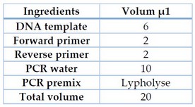
Table 1. PCR premix master mix Volume
PCR tubes were prepared according to manufacture instructions, then transferred into spin and vortex centrifuge at 300 rpm for 3 minutes at 3000 rpm, then placed in PCRThremocycler PCR complication condition shown in Table 2
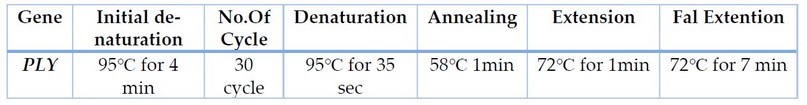
Table 2. PCR condition
PCR products were resolved on 1% agarose gel, as shown by UV illumination and photographed 14.
Sequence analysis
Ply PCR products were sent to Macrogene Korea for sanger sequencing.
Comprehensive phylogenetic tree
The evolutionary history was inferred using the UPGMA method 15. The optimal tree with the sum of branch length = 6.56300659 was shown. The tree was drawn to scale, with branch lengths in the same units as those of the evolutionary distances used to infer the phylogenetic tree. The evolutionary distances were computed using the Maximum Composite Likelihood method 16 and are in the units of the number of base substitutions per site. The analysis involved 19 nucleotide sequences. Codon positions included were 1st+2nd+3rd+Noncoding. All positions containing gaps and missing data were eliminated. The ply sequencing was compared with similar sequences using NCBI-BLAST (https://www.ncbi.nlm.nih.gov/). There was a total of 200 positions in the final dataset. Evolutionary analyses were conducted in MEGA6, and nucleotide and amino acids were analyzed in Genes program 17.
RESULTS
Isolation and Identification of Streptococcus pneumoniae
Microscopic examination of isolates showed gram-positive diplococci lancet-shaped cells Figure 1. In contrast, on blood agar, the isolates exhibited α-hemolysis Figure 2 as well it was susceptible to optochin when submitted to the optochin susceptibility test Figure 3.
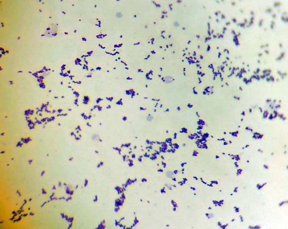
Figure 1. S.pneumoniae under the light microscope (100X)
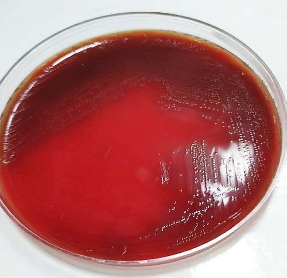
Figure 2. S.pneumoniae on blood agar
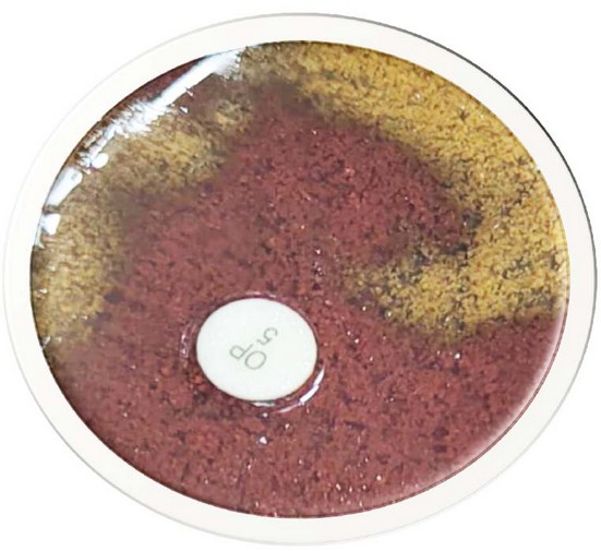
Figure 3. Optochin sensitivity test of S.pneumoniae
Result of investigation on pneumolysin
The results of the investigation of pneumolysin by the agar double layer method showed that S.pneumoniae has the ability to produce pneumolysin, as no growth was appeared on the surface of the culture medium after inoculation with bacterial types which were sensitive to pneumolysin including gram positive bacteria S.aureus figure 4 and gram negative bacteria E.coli figure 5.
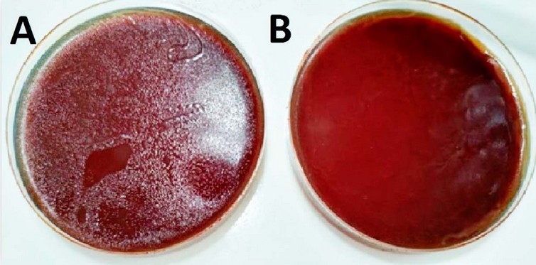
Figure 4. E.coli susceptibility to pneumolysin. A: control plate. B: test plate
Investigation of ply gene by using PCR technique
DNA primers amplify a specific ply gene by polymerase chain reaction (PCR). DNA was extracted, and using a Nanodrop device, the concentration of DNA was measured, which ranged between (51-112 ng/μ1), and its purity was determined with (1.8-2). The ply gene was amplified to the size of 238 base pairs compared to the DNA ladder 100bp, and the result matched with the standard isolate Streptococcus pneumoniae ATCC 49619. The results showed that 3 isolates from 5 (60%) contained ply gene, as in Figure 5.
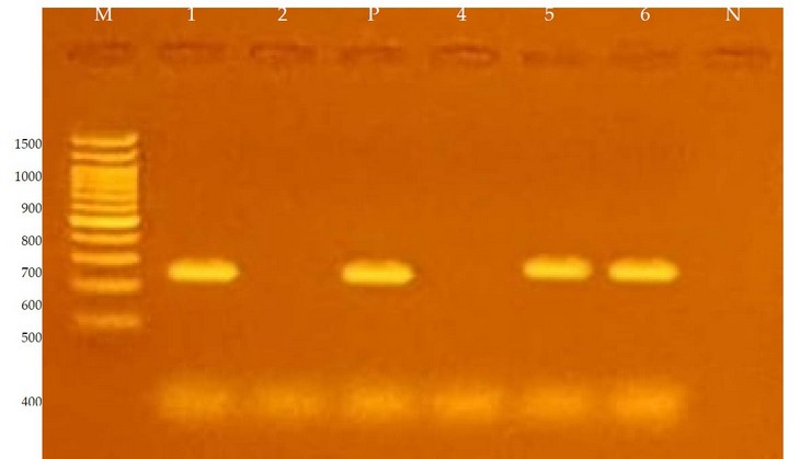
Figure 5. Electrophoresis for amplification products of ply in a 1% agarose gel with 65 volts and for an hour and a half using M path
DNA ladder starts from 100 base pairs, the path N represents the negative control, P represents positive control stander isolate Streptococcus pneumoniae ATCC 49619, columns representing the Gene ply of the Streptococcus pneumoniae isolates and their size 238 base pairs.
. ply sequencing
Ply sequences were obtained by Sanger sequencing
Genetic variation of ply gene
Allele of ply (n=7) was identified compared to 18 known ply alleles. Three amino acid changes gave three mutations, Table 3. The Allele subtype found that nucleotide sequences varied more than amino acid sequences.
The identity was high in alleles based on the nucleotide and amino acid sequences using pairwise comparisons of our isolate and EF413936.1; the identities were 99%. Our isolate had genetic variation at the ply loci allele (n=7). This locus may be replaced with foreign DNA through gene transfer mechanisms like transformation, conjugation, transduction and lysogenic conversion. This mutation changes the amino acid arginine to valine, a missense mutation.
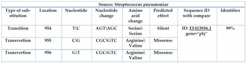
Table 3. Nucleotide and amino acid change with location
Transvertion
The local isolate showed a 99% match with the 17 globally recorded isolates while showing a 94% match with standard isolate S.pneumoniae 49619 ATCC fig (6)
Phylogenetic analysis
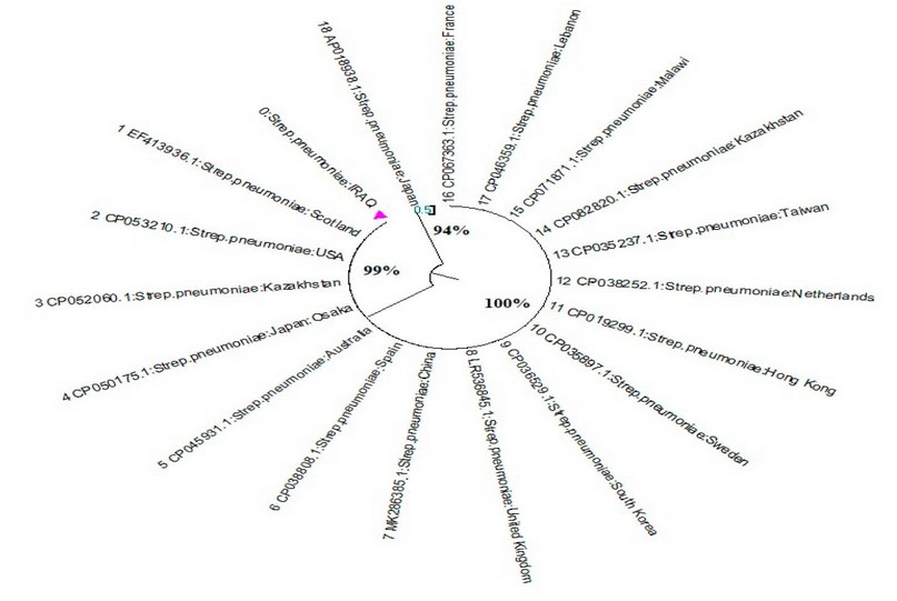
Figure 6. Phylogenetic tree of the ply variants of S.pneumoniae isolates
DISCUSSION
Five Streptococcus pneumoniae isolates were obtained from 50 sputum specimens with a 10% percentage. This percentage was lesser than the percentage obtained by Tamimi Zainab 18 where the ratio of isolation of this bacterium was 11%, but higher than the isolation rate of the study of 19, who found the isolation rate was 4%. The inhibitory activity of pneumolysin produced by S.pneumoniae on both gram-positive S.aureus and gram-negative E.coli were agreed with 20 who used the same bacterial types that were sensitive to pneumolysin gram-positive S.aureus and gram-negative E.coli. Nucleotides 954,955,956 were identified with position variation, (T954C),(C955G),(G956T) substitution replacement mutation in (T954C) without effect on encoding amino acid there for named silent) because the two codons (AGT) and (AGC) encode to Serine amino acid and no phenotypic will be changed as a result of this replacement, but the two-locus,(C955G),(G956T) were shown to affect encoding amino acid Arginine, nucleotide changes lead to amino acid change; as a result to change with the start codon and instead of encoding to produce Arginine will coding to Valine 21.
CONCLUSION
Due to the role of pneumolysin, which represents a key for infection with S.pneumoniae so this study aimed to highlight on isolation and identification of Streptococcus pneumoniae using morphological, biochemical and Vitek, as well as investigation of pneumolysin phenotypically and molecularly through ply gene and sent the PCR products to sequencing by sanger method.
REFERENCES
1. Brooks, L. R., &Mias, G. I. Streptococcus pneumonia’s virulence and host immunity: aging, diagnostics, and prevention. Frontiers in immunology, 2018, 9, 1366.
2. Jia, J., Shi, W., Dong, F., Meng, Q., Yuan, L., Chen, C., & Yao, K. Identification and molecular epidemiology of routinely determined Streptococcus pneumoniae with negative Quellung reaction results. Journal of Clinical Laboratory Analysis, 2022; e24293.
3. Ali, A. F. .; Mohammed, T. T. .; Al-Bandar, L. K. . The Role Of Optifeed®, Vêo® Premium, And Oleobiotec® In Diets For Improvement Of The Production Performance Of Male Broilers In Heat Stress. JLSAR 2022, 3, 32-36..
4. Tong, N., & BA, M. Background paper 6.22 pneumonia. A Public Health Approach to Innovation, 2013; 1(1), 7-8.
5. Kurihara, E., Takeshita, K., Tanaka, S., Takeuchi, N., Ohkusu, M., Hishiki, H., &Ishiwada, N. Clinical and Bacteriological Analysis of Pediatric Pneumococcal Meningitis after 13-Valent Pneumococcal Conjugate Vaccine Introduction in Japan. Microbiology Spectrum, 2022; e01822-21.
6. Silbergleit, M., Vasquez, A. A., Miller, C. J., Sun, J., & Kato, I. Oral and intestinal bacterial exotoxins: Potential linked to carcinogenesis. Progress in molecular biology and translational science, 2020; 171, 131-193.
7. Garvey, M.; Rowan, N. J. and Pulsed, U. V. As a potential surface sanitizer in food production processes to ensure consumer safety. Curr. Opin. Food Sci. 2019; 26: 65- 70. doi: 10-1016/j.ijbiomac. 2018.05.233.
8. Ding, R., Zhang, Y., Xu, X., Hou, Y., Nie, J., Deng, X., ...&Lv, Q. Inhibitory effect of hederagenin on Streptococcus pneumoniaepneumolysin in vitro. Microbes and infection, 2022;24(2), 104888.
9. Vögele, M., Bhaskara, R. M., Mulvihill, E., van Pee, K., Yildiz, Ö.,Kühlbrandt, W., ... & Hummer, G. Membrane perforation by the pore-forming toxin pneumolysin. Proceedings of the National Academy of Sciences, 2019;116(27), 13352-13357.
10. Holt J G, Krieg N R, Sneath P H A, Staley J T and Williams S T. Bergey’s manual of determinative bacteriology. 9th ed. Williams & Wilkins: Baltimore, Maryland; 1994; pp. 20 & 527-558 .
11. Al-Bazy, F. I. .; Abdulateef, S. M. .; Sulimn, B. F. . Impact Of Feeds Containing Optifeed®, Vêo® Premium, And Oleobiotec® On The Lipid Peroxidation Of Male Broilers Under Heat Stress. JLSAR 2022, 3, 25-31..
12. Hockett, K. L., &Baltrus, D. A. Use of the soft-agar overlay technique to screen for bacterially produced inhibitory compounds. JoVE (Journal of Visualized Experiments), 2017 ;119, e55064.
13. www.ncbi.nlm.nih.gov (https://www.ncbi.nlm.nih.gov/)
14. Han, C., & Zhang, M. Genetic diversity and antigenicity analysis of Streptococcus pneumoniaepneumolysin isolated from children with pneumococcal infection. International Journal of Infectious Diseases, 2019; 86, 57-64.
15. Sneath P.H.A. and Sokal R.R. (1973). Numerical Taxonomy. Freeman, San Francisco.
16. Alkubaisy,S.A., A.A. Majid, S.M. Abdulateef, F.A. Al-Bazy, O.K. Attallah, O.M. Abdualmajeed, Th. T. Mohammed, F.M. Abdulateef, K.I. Mahmud. Effects of In-Ovo injection of Biotin on chick's embryonic development and physiological traits. IOP Conference Series: Earth and Environmental Science, 2021; 761(1), 012111.
17. Tamura K., Stecher G., Peterson D., Filipski A., and Kumar S. MEGA6: Molecular Evolutionary Genetics Analysis version 6.0. Molecular Biology and Evolution, 2013; 30: 2725-2729.
18. Tamimi Zainab Amer Hatem. Bacteriological and hereditary study of Streptococcus pyogenes isolated from patients with tonsillitis in the city of Muqdadiya, Master Thesis, College of Education for Pure Sciences \ University of Diyala. 2013.
19. Keith, E. R., Podmore, R. G., Anderson, T. P., & Murdoch, D. R. Characteristics of Streptococcus pseudopneumoniae isolated from purulent sputum samples. Journal of clinical microbiology, 2006; 44(3), 923-927.
20. Yan, H., Lu, Y., Li, X., Yi, Y., Wang, X., Shan, Y., ...&Lü, X. Action mode of bacteriocin BM1829 against Escherichia coli and Staphylococcus aureus. Food Bioscience, 2021; 39, 100794.
21. Schenk, M. F., Zwart, M. P., Hwang, S., Ruelens, P., Severing, E., Krug, J., & De Visser, J. A. G. M. Population size mediates the contribution of high-rate and large-benefit mutations to parallel evolution. Nature Ecology & Evolution, 2022; 1-9.
Received: May 15, 2023/ Accepted: June 10, 2023 / Published: June 15, 2023
Citation: Thamer, R.A.; Al-Rawi, A.M. Phenotypic and molecular Investigation of Streptococcus pneumoniae pneumolysin. Revis Bionatura 2023;8 (2) 57. http://dx.doi.org/10.21931/RB/2023.08.02.57