2022.07.02.58
Files > Volume 7 > Vol 7 No 2 2022

Noor Hussein Yousif
Iraq Natural History Research Center &Museum \ University of Baghdad. Iraq
Corresponding author: [email protected]
Available from: http://dx.doi.org/10.21931/RB/2022.07.02.58
ABSTRACT
Bones were recorded in the skeleton of some species of Iraqi turtle Mauremys rivulata; the objectives of this study came in light of current conditions, environmental developments, talents and techniques of biological studies taking place in the country, need for an anatomy guide in river turtles of Iraqi species, to identify all kinds of similarities and differences with their preaching, this work or study has become written in response to those modern needs. It is designed to be one of the resources for those interested in biological studies, beginners or professionals, and veterinarians, distinguishing them from marine and global species. Turtles were dissected in the laboratories of the Research Center and Museum of Natural History / the University of Baghdad. The specimen was dissected by removing the abdominal cortex, muscles, and internal viscera and imaging the bone starting from the skull to the hind leg bones. This first study was in Iraq.
Keywords: Turtles (Mauremys rivulata), anatomy, skeleton, bone, Iraq
INTRODUCTION
The history of turtles(Mauremys rivulata) goes back to the Jurassic period when they were found about 210 million years ago. They are considered the oldest primitive armored animal with some standard features with the others to this day, and the most prominent thing in common is a bony shell that consists of a group of ribs that fuse to form that apparent solid plate1. Turtles are classified into two main types: 1- pleurodires that bend to one side for each head and neck. 2- Cryptodires that pull the head back in a vertical and straight line between their shoulders is known as the movement of the letter S 2. These river turtles produced many sea turtles that trace their origins to the Cretaceous period, about 65 million years ago. Based on fossils found in different regions of the world, 3.
Mauremys rivulata is a turtle that descends from the coasts of Croatia, Albania, and Macedonia with Greece and part of southwestern Turkey, through the Middle East, Iraq, Jordan, and Syria, Palestine, and Lebanon4. Its presence in these places is due to the weather and the moderate environment that suits it5. M. rivulata in static and slow waters such as lakes, irrigation streams, swamps, estuaries, ponds, and even dams are the habitats you prefer to be in rather than fast-flowing water6,7
The turtle's body outline is classically represented as a result of morphology and development, as it has a dorsal shield and also there is ossified abdominal support with four legs, two front and two hinds; the nature and shapes of the turtle shields are still under the interest of scientists, the shield is a part of the axial cartilaginous skeleton of the thoracic vertebrae and the ribs with a group. The dermis that overlaps with it, and there are many dermis forms, magnifies its component in this great mosaic8,9.
The ossification of the skeleton in the skull is typical of vertebrates. However, it is rare after the cranium that the ribs change according to the limbs and the girdles between them, the abdominal or thoracic brace interferes with the thoracic or pelvic girdles in the front and backside of the ribs due to its close association with the dermis. To maintain the position Dorsal, tetrapods are related to the axis of the dorsal girdle ventral deep; in this study, the bony parts will be clarified10.
Some studies author study the evolutionary inclusion of the turtle's body and how it evolved from the primitive tetrapods to the more typical shape as a result of a developmental conflict between similarity to the structural elements in turtles11,12.
The skeleton of sea turtles is characterized by heavyweights that reach about a (1ton) and a length of about 2 meters13. Also consists of the dorsal and abdominal supports, axial skeleton, and four legs. The number of bones was found up to (280) bones of different shapes and sizes14,15,16.
Turtles have been exploited over the years as they are considered a nutritional value for some coastal areas; the turtle shell has been used in various cultures 17,18,19. In comparison, the bones do not attract scientists much attention to their relatively large number with the diversity of their shapes and sizes for the prominent types of turtles with the recording of numbers of bones In some records of archaeological areas18,19. The movement of the spine is an essential requirement for every animal. The reason for movement is not to obtain food but because it needs movement to escape predators20. The turtle shell is an excellent shield for it because it contributes to protecting the head and neck and the internal viscera, and the head retracts vertically or horizontally laterally inside the shell 21. A turtle's neck consists of eight cervical vertebrae articulated with the skull with a group of complex joints that facilitate movement 22.
Classification of turtle
· Kingdom: Animalia
· Phylum: Chordata
· Class: Reptilia
· Order: Testudines
· Suborder: Cryptodira
· Superfamily: Testudinoidea
· Family: Geoemydidae
· Genus: Mauremys
· Species: M. rivulata
· Binomial name 23,24.
MATERIALS AND METHODS
Samples were taken from irrigation creek water from Babel and Diyala provinces during the summer season (June- July 2021 ). Four species of adult turtles Mauremysrivulata (2 ♂, 2 ♀), male weight 660 ± 3.30 g and length of 25 ± 1.39 cm and female weight 560 ± 2.30g and length of 23 ± 1.24 cm, The animal was anesthetized, and then the abdominal shell was removed with anatomical tools. The internal viscera was removed, all parts of the shell and bones were cleaned from the remains of muscles, the bones were extracted for the front and back legs with the head and tail and studied in terms of shape and location and compared to each other 25.
RESULTS
The shell
At the beginning of the study, the female had shells darker in color than the males. The dorsal shells have 40 in number and the ventral ones have12 in number, while the dorsal shells of the males have 37 and the ventral ones have 11, according to (Fig.1) ., and from here, it was found that the females are more comprehensive to they carry eggs inside them.
The shells were fused to the turtle's spine with a trace of the ribs that may have been fused with the shells for additional protection and support for the animal from dangers as a defense mechanism (Fig.2).
The skull and vertebrae
Is unpretentious in composition, consisting of the temporal bone fused with the orbit of the eye socket and the nasal bone opening. Additionally, the two ear holes and jaw bone discriminate between the upper and lower mandible containing the teeth (Fig.3). The 6 cervical vertebrae are fused with the skull; the straightforward form is fused. The last vertebra is fused with the sternum, collarbone, and dorsal shell. (Fig.4). thoracic vertebrae 8 in number is, fused with the shells. The remains of the ribs and shells are shown in (Fig.5).
The lumbar vertebrae are 4 in a number smaller than the thoracic vertebrae. They also fuse with the dorsal shell. Furthermore, the sacral or coccygeal vertebrae show 3vertebrae fused to form one bone fused with the dorsal shell (Fig.6).
The caudal vertebrae in females were smaller in size than in males. They contain the shape of the vertebrae in the advanced vertebral organism. Their number ranges from 20-22 vertebrae that are not fused with the shells. (Fig.7).
Appendicular skeleton
Forelimbs, the scapula is a flat bone showed that does not contain the protrusion of the scapula and articulation with the humerus bone, which was more prominent in males than females, and the presence of the radius and ulna; these two bones were relative in shape to each other, but the radius bone was thicker than the ulna (Figs 6,8, and 9).
It articulates with the carpal bone, which is 10 tiny bones in each of the anterior vertebrae, fused and articulating with the phalanges (Fig.9).
The fingers in both sexes were 5 in the front limbs and 4 in the hind limbs, which were longer than the front fingers and contained claws, hind limbs, The femur bone, which articulates with the pelvic bone on the proximal side, and articulates with the tibia and fibula on the distal side, the tibia, and fibula, in two sexes tibia thicker than fibula but male thicker than female and those two bones. Articulation with 5 small metacarpal bones fused together and connected with phalanges and fingers (Fig.10 and 11).
The pelvic bone in females is more comprehensive than in males due to the presence of the ovary and the female reproductive organs. It contains three fused bones that make up the pelvis: ischium, ilium, and pubis. (Fig. 7).

Figure 1. show dorsal and ventral (a) male and (b)female shall

Figure 2. Show ribs fused with shall in (a) male and (b) female shall
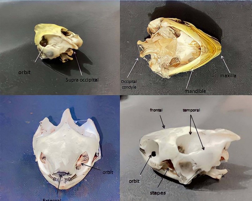
Figure 3. Show skull in the two species the same

Figure 4. Show the occipital condyle articulation with 1st cervical vertebrae
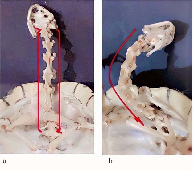
Figure 5. show cervical vertebrae (a) male (b)female

Figure 6. The black arch show thoracic vertebrae fused with ribs and shall ,red circle the pelvic bone (ileum +ischium ,pubis , red arrow its scapula ,black arrow the clavicle bone blur arrow the coracoids bone

Figure 7 . Show caudal vertebrae And pelvic bone (ischium, ilium, and pubis) (a) male (b) female

Figure 8. Show forelamb red arrow humerus bone ,black head arrow head of humerus ,brown arrow scapula and blue arrow the clavicle bone .
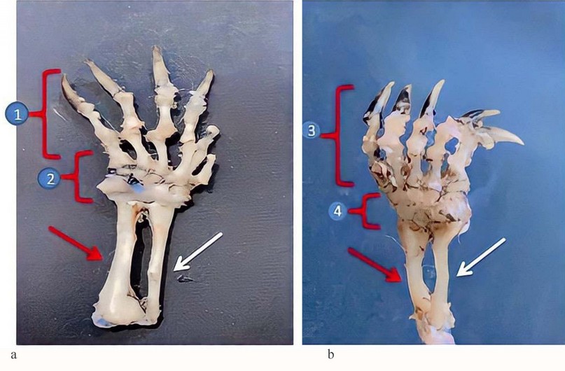
Figure 9. shows forelimb red arrow ulna bone , white arrow radius bone, 1&3– phalanges 2&4- carpal bone

Figure 10. Show hind lamb red arrow femur bone, black arrow tibia bone, blue arrow fibula bone.

1- Phalanges
2- Metacarpal (tarsal) bone
Figure 11. Red arrow tibia bone, black arrow fibula bone.
DISCUSSION
Auther used almost all sea turtles - including marine ones belonging to the family Cheloniidae –to have a shell consisting of a dorsal and ventral shield, and the material that does not consist of it is carnelian brown in color26. Like river turtles with different colors, they tend to the dorsal shell to dark brown color in males and females, while the shell The abdomen is dark green. The name of the tortoiseshell in the literature came (ar chae o log i cal and eth no Graph ic). It is an unclear term referring to the shell in the dorsal and ventral parts, considered a carotenoid shell or bony horn. In many cases, this is clear from context 27. The osteology section of this atlas is The bones of the skull (prefrontal, frontal, parietal, postorbital, supraoccipital, squamosal, quadratojugal, jugal, and maxilla) and mandible (dentary, angular, surangular, prearticular, splenial, and articular bones) while.
The results observed that the skull had undifferentiated fused bones consisting of the temporal bone fused with the orbit of the eye socket and the nasal bone opening, the two ear holes, and the discrimination of the upper and lower mandible containing the teeth28. The observed author explained that the neck is long for evasion and capture of prey, consisting of cervical vertebrae that help it to move freely, crouching inside the body when sleeping and feeling fear, comparable to what was explained29,30. The neck is long in all species, whether aquatic or semi-aquatic. Control of the neck is crucial for turtles as it is multi-joint with a complex structure consisting of developed vertebrae to the neck, body, and head of the vertebra are articulated to make them more accurately and efficiently and control their freedom during movement during rapid movements in proportion to Its nature, and this is indicated by 22 where this development of the cervical vertebrae is proportional to the accompanying muscles that allow them to move vertically or horizontally river turtles in Iraq are little studied. However, cervical vertebrae were found in all types of turtles that supply eight rectangular vertebrae (C1 to C8) and nine joints. observed31.
Observed appendicular skeleton in their study, there were some similarities in the skeleton of the thoracic and pelvic girdle and the fore and hind limbs in the reptiles studied; P. guttatus and A. boskianus32. With the presence of incommodious differences in the length of the angled ends of the bones in the thoracic and pelvic, which is similar at the same time to river turtles with fewer bones in size and thickness, a similarity was found in terms of the presence of double wishbones on the non-double-bone river turtles are more developed than them 33. The skeleton of the forelimbs of the same studied, which consists of the humerus, radius, ulna, and metacarpal bones, was observed, and it is similar to the river turtles studied in this research, and it is connected with the thoracic girdle, and in general, it is identical to mammals and birds The bone radius was a long bone articulated with the carpal uncle and ulna as in turtles, while the ulna was more prominent and thicker in sources, unlike river turtles, it was a slight difference in thickness. Studies of reptiles assured P. guttatus and A. boskianus having a pelvic girdle consisting of the pubic, iliac, and ischium; It fused and met the acetabulum, and they left a wide foramen (fenestra ischio-pubic fenestra). There was a clear difference in the shape of the pubis, ischium, and iliac of the two species studied with river turtles in terms of size and thickness with pelvic bones32,34.
The study contained the tibia and fibula of approximately equal length but shorter and thinner than the femur and also noted that the tibia is more straight than the fibula and articulated with the femur by the tibial condyle of the femur. The distal part is articulated with the heel bone and is analogous to the study of a developed vertebrate35.
The phalanges and their total number in the anterior and posterior extremities, respectively (manus: 2-3-3-3-2, pes: 2-3-3-3-1). It is similar to river turtles except that the back end contains five digits, only in C. flavomarginata, it is also found with a missing phalanx in the 5 digit of manus and pes; They also appear in species in the pes of C. mouhotii. In C. flavomarginata, the fifth digit of pes is also missing. Due to changes in some other terrestrial and semi-terrestrial geological materials (Cuora mccordi, Heosemys spinosa, part C. mouhotii and Leucocephalon yuwonoi) they also took a similar pattern which led to the disappearance of the fifth finger of pes. It is also found in turtles (Testudinidae), and in another group of terrestrial Chelonians, and land turtles of the genus Terrapene (Emydidae); missing phalanxes or whole digits are known to be found in the species Malayemys subtrijuga, Morenia petersi, Pangshura smithii, and Siebenrocki that have been studied where The finger found the fifth digit of the manus(2-3-3-3-3); on one side of the body there are phalanxes of P. 3-3) These are aquatic turtles came similar to the study of river turtles36.
Since there are few studies of river turtles, the writer discussed the jaw with reptiles, and as it is known, their lower jaw consists of several bones. Cuvier gave them a descriptive name in line with the jaws of crocodiles, turtles, lizards, and snakes in the order named with slight differences in turtles
In 37 the wishbone was not distinct or was small in the thoracic girdle, while in river turtles, the wishbone is found clearly and distinctly. It has thick and convex borders.
CONCLUSION
This study observed the skeleton of Iraqi river turtles in general and is considered the first study in Iraq. The simplicity of the composition concerning the skull is obvious, with the development of the limbs and their differentiation and difference from sea turtles in the presence of claws, and it required a separate study for each part of the skeleton.
REFERENCES
1- Gaffney, Eugene S. The comparative osteology of the Triassic turtle Proganochelys. Bulletin of the AMNH ,1990; no. 194
2- Perrine, DSea Turtles of the World. Voyageur Press, Stillwater, Minnesota, 2003.
3- Cameron, C. Canadian Canal Society fonds, 1896, 1904, 1966, 1973-2013.
4- LC, N., NT, N. and NE, N., Turtle Taxonomy Working Group (van Dijk, PP, JB Iverson, AGJ Rhodin, HB Shaffer, and R. Bour). Turtles of the world: annotated checklist of taxonomy, synonymy, distribution with maps, and conservation status. Chelonian Research Monographs, 2014; 5 (7): 329-479.
5- Fritz, U., & Wischuf, T. Zur Systematik westasiatischsüdosteuropäischer Bachschildkröten (Gattung Mauremys). Zoologische Abhandlungen, Staatliches Museum für Tierkunde Dresden, 1997; 49: 223–260
6- Jones MEH, Werneburg I, Curtis N, Penrose R, O'Higgins P, et al. The Head and Neck Anatomy of Sea Turtles (Cryptodira: Chelonioidea) and Skull,2012.
7- Mantziou, G. and Rifai, L. Mauremys rivulata (Valenciennes in Bory de Saint-Vincent 1833)—Western Caspian Turtle, Balkan Terrapin. Conservation Biology of Freshwater Turtles and Tortoises: A compilation Project of the IUCN/SSC Tortoise and Freshwater Turtle Specialist Group, Chelonian Research Monographs, 2014; 5: 080-1.
8- Owen, R. On the development and the homol- ogies of the carapace and plastron of the chelonian reptiles. Phil. Trans. R. Soc. London (Biol.) ,1849;151-171.
9- Zangerl, R.. The turtle shell. In C. Gans and A. d'A. Bellairs (eds.), Biology of the Reptilia,. Academic Press, London,1969 ;1: 311-339
10- Billett, F., C. Gans, and P. F. A. Maderson. Why study reptilian development? In C. Gans, F. Billet, and P. F. A. Maderson (eds.), Biology of the Reptilia, 1985; 14: 1-40.
11- Burke, A.C. The development and evolution of the turtle body plan: inferring intrinsic aspects of the evolutionary process from experimental embryology. American Zoologist, 1991;31(4):616-627.
12- Burke, A. C. Origin of the Turtle Body Plan. Great Transformations in Vertebrate Evolution, 2015;77-89
13- Pritchard, P. C. H. & Mortimer, J. A. Taxonomy, external morphology, and species identifi cation. K. L. Eckert, K. A. Bjorndal, F. A. Abreu-Grobois & M. Donnelly (eds.). Research and Management Techniques for the Conservation of Sea Turtles. IUCN/SSC Marine Turtle Specialist Group Publication,1999; 4: 21–38.
14- Mosseri-Marlio, C. Sea turtle and dolphin remains from Ra's al-Hadd, Oman. – M. Mashkour, A. M. Choyke, H. Buitenhuis & F. Poplin (eds.). Archaeozoology of the Near East, IVB. ARC-Publicatie, 32. Groningen, 2000b; 94–103.
15-Mosseri-Marlio, C. Sea turtles and dolphins: Aspects of marine animal exploitation from Bronze Age Rā's al Hadd, Oman. – The Journal of Oman Studies, 2002;12: 197–210.
16- Wyneken, J. The Anatomy of Sea Turtles. U.S. Department of Commerce, National Oceanic and Atmospheric Administration, National Marine Fisheries Service, Southeast Fisheries Science Center, Miami, Florida; NOAA Technical Memorandum NMFS-SEFSC-471, 2001.
17- Thode-Arora, H. Tapa und Tiki. Die Polynesian-sammlung des Rautenstrauch-Joest-Museums. Ethnologica Neue Folge, 23. Rautenstrauch-Joest-Museums der Stadt Köln. Köln,2001.
18- Frazier, J. Prehistoric and ancient historic interactions between humans and Marine turtles. – P. L. Lutz, J. A. Musick & J. Wyneken (eds.). The Biology of Sea Turtles, 2. CRC Press. Boca Raton, Florida, 2003; 2: 1–38.
19- Frazier, J. Marine turtles of the past: A vision for the future? – R. C. G. M. Lauwerier & I. Plug (eds.). The Future from the Past: Archaeozoology in Wildlife Conservation and Heritage Management. Proceedings of the 9th ICAZ Conference, Durham 2002, 3. Oxbow Books. Oxford, 2004:103–116.
20- Irschick, D.J., and T. Garland, Jr. Integrating function and ecology in studies of adaptation: investigations of locomotor capacity as a model system. Annual Review of Ecology and Systematics ,2001; 32:367–396.
21- Peumans, W. J., & Van Damme, E. J. Lectins as plant defense proteins. Plant physiology, 1995;109(2): 347.
22- Aerts, P., Van Damme, J., & Herrel, A. Intrinsic mechanics and control of fast cranio-cervical movements in aquatic feeding turtles. American Zoologist, 2001; 41(6), 1299-1310.
23 - Corduk, N., Dogru, N. H., Gull, C., & Tosunoglu, M. Assessment of nuclear abnormalities in erythrocytes of balkan pond turtle Mauremys rivulata (Valenciennes, 1833)(Testudines: Geoemydidae) from the Biga Stream, Çanakkale, Turkey, 2019.
24 -Bayrakcý, y., Ayaz, d., Yakýn, b.y., Çiçek, k. and Tok, c.v. Abundance of western caspian turtle, mauremys rivulata (valenciennes, 1833) in gökçeada (imbros), turkey. russian journal of herpetology, 2016; 23(4).
25- Tompestt, D.H. Anatomical techniques 2nd add. Longman group limited. London Great Britain, 1970.
26- Benscoter, Allison M.; Smith, Brian J.; Hart, Kristen M. Loggerhead marine turtles (Caretta caretta) nesting at smaller sizes than expected in the Gulf of Mexico: Implications for turtle behavior, population dynamics, and conservation. Conservation Science and Practice, 2022; 4.1: e581.
27- Rudrud, Regina Woodrom. Forbidden sea turtles: traditional laws pertaining to sea turtle consumption in Polynesia (including the Polynesian outliers). Conservation and Society, 2010; 8(1): 84-97.
28- Arencibia A, Melián A, Orós J. Anatomic Interactive Atlas of the Loggerhead Sea Turtle (Caretta caretta) Head. Animals , 2021; 11(1):198.
29- Herrel, A., Van Damme, J., & Aerts, P. Cervical anatomy and function in turtles. In Biology of turtles CRC Press, 2007; 177-200.
30- PRITCHARD, Jack A.; CUNNINGHAM, F. Gary; PRITCHARD, Signe A. The Parkland Memorial Hospital protocol for treatment of eclampsia: evaluation of 245 cases. American journal of obstetrics and gynecology, 1984;148(7): 951-963.
31- Fritz, U., Auer, M., Bertolero, A., Cheylan, M., Fattizzo, T., Hundsdörfer, A.K., Martín Sampayo, M., Pretus, J.L., ŠIrokÝ, P. and Wink, M., A rangewide phylogeography of Hermann's tortoise, Testudo hermanni (Reptilia: Testudines: Testudinidae): implications for taxonomy. Zoologica Scripta, 2006; 35(5):531-543.
32- Ali, W., Javid, A., Hussain, A., & Bukhari, S. M. Diversity and conservation of freshwater turtles in Pakistan: a review. Biodiversity, 2018;19(1-2):62-71.
33- Zaff A, Herrel A, Aerts P and De Vree F .Morphology and morphometrics of the appendicular musculature in geckos with different locomotor habitat. Zool. Morphol., 1999;119: 9-22.
34- Saber S A, ElSalkh B A, Gadel-Rab A G, Mahmoud F A, El Dahshan A A and Gewily D I. Comparative and functional study of integumentary system of two different reptiles: adaptation to their different modes of life. The Egyptian J. of Hospital Medicine, 2018; 73 (6): 6802-6811.
35- Gadel-Rab, A. G., Mahmoud, F. A., Saber, S. A., ElSalkh, B. A., El-Dahshan, A. A., & Gewily, D. I. Comparative Functional Analysis of the Anatomy of the appendicular skeleton in two reptilian species. The Egyptian Journal of Hospital Medicine, 2018; 73(8), 7274-7287.
36- Fritz, U., Auer, M., & Petzold, A. Osteology in the Cuora galbinifrons complex suggests conspecifity of C. bourreti and C. galbinifrons, with notes on shell osteology and phalangeal formulae within the Geoemydidae, Amphibia-Reptilia, 2006; 27(2), 195-205.
37- Williston, S. W. The osteology of some American Permian vertebrates. The Journal of Geology, 1914; 22(4): 364-419.
Received: 21 February 2022 / Accepted: 31 March 2022 / Published:15 May 2022
Citation. Hussein Yousif N. Comparative anatomical study to skeleton for same species of Turtles in Iraq. Revis Bionatura 2022;7(2) 58. http://dx.doi.org/10.21931/RB/2022.07.02.58