2022.07.04.2
Files > Volume 7 > Vol 7 No 4 2022
Determination of IFN-y in Patients with Pseudomonas aeruginosa-Inflicted Burn and Wound
Mohammed A. Jwad 1, Wasan A. Gharbi 2
* Correspondence: [email protected]; Tel.: (07737742737)
Available from: http://dx.doi.org/10.21931/RB/2022.07.04.2
ABSTRACT
The sample collection was carried out from 1/12/2021 to 1/4/2022 at the burns hospital, Baghdad teaching hospital in Baghdad city, and Alhussain teaching hospital in Almuthana city. Samples were collected from patients' sera collection samples from (69) patients, from burn patients 53 and 16 samples from wound patients infected with pseudomonas aeruginosa; 41 patients were males, and 28 were females. This group used the other 69 patients that were not infected by bacteria as a control group. The level of IFN-y was investigated by ELISA assay in the teaching Laboratories in al Sawawah city. The results showed a decrease in IFN-y levels were 35.2±2.6 for burn patients and 35.4±2.3 for wound patients compared to the control group of 46±2.5. The current study reported a highly significant difference in IFN-y levels between the burn and wound patients and the control group (P<0.0001 ).
Keywords: Pseudomonas aeruginosa; Burn; wound; Interferon – Gamma.
INTRODUCTION
Pseudomonas aeruginosa is a common pathogenic germ that can cause significant opportunistic infections, especially in immunocompromised people. The spread of this organism in healthcare facilities is highly harmful, as it infiltrates the human host's primary defensive line and enters the body through the skin, resulting in nosocomial infections, particularly in hospital intensive care units (ICUs). Due to the availability of various mechanisms of natural resistance to most antibiotics, the pathogenesis of P.aeruginosa is multifactorial, resulting in the creation of a diverse set of cellular structures and extracellular chemicals that play a crucial role in increasing pathogenicity 1 A rod-shaped GN bacteria P.aeruginosa is a common cause for hospital-acquired illnesses. While HPV usually does not affect healthy people, it can colonize any part of the human body that has enough moisture to form a niche. P. aeruginosa is responsible for roughly 8-10% of all healthcare-associated infections in the United States (51,000 cases in 2013). Burns is one of the most common types of trauma destruction 2.
Burns compromise skin integrity and the skin's immune system, which protects against pathogenic organisms' activity 3. Nosocomial infection is a significant concern for burn patients. Infection is a primary source of morbidity and mortality in hospital burn patients. Because of their weakened state and the nature of the damage, nosocomial infection is more common in burn patients 4 Wound, by definition, breaks in skin epithelial integrity and may cause further disruption in skin anatomy, physiology, and functions. There are two types of wounds known as acute and chronic. 5 In response to harmful agents such as bacteria, tumor cells, viruses, and parasites, vertebral cells produce interferon-gamma (IFN-y). IFN-y is a critical cytokine in adaptive and innate immunity to intracellular microorganisms. IFN-y increases T and B cell development, activates macrophage microbicidal activity, and boosts cytotoxic T cells. 4
MATERIALS AND METHODS
Species study
The present work includes 138 the collection of serum from (53) burn patients and (16) wound patients that infection with pseudomonas aeruginosa; 41 patients were males, and 28 were females; the other 69 patients were not used patients not infected by bacteria as a control group. The samples were collected from burns hospitals in Baghdad and Almuthanna city from 1/12/2021 to 1/4/2022. These samples were used to investigate the IFN-y in the patients and control group. The sera were collected, brought to the teaching laboratories in Almuthanna and tested. This study used the IFN-y kits to perform the assay using the ELISA instrument. The company that accoutered the kits was Shanghai YL Biont / China.
.
Sample Collection and Storage
10mls of venous blood was carefully drawn into appropriate sample bottles and spun to separate the serum. The serum was separated into a plain sterile sample bottle and stored at -20o C for analysis.
Data Management and Analysis
The study was designed by a Completely randomized design (CRD) that was used in the analysis of variance for data of gamma interferon values by using a one-way ANOVA test, independent t-test, and Dunnett's test at a 5% level of significance. Moreover, All frequency data were analyzed by Pearson's chi-squared test and Fisher's exact test. Data were processed and analyzed by using statistical program social science (SPSS 22), and the results were expressed as Mean±SD or percentages 6
Laboratory Procedure
In vitro test for the quantitative determination of ELISA serum gamma interferon . The blood was drawn through a syringe, added to a gel tube, then transferred to a centrifuge to separate the blood components from the serum, and examined in a machine on ELISA system and Elisys uno Germany gamma interferon Test.
Assay procedure
Standard solutions: (This kit has a standard original concentration, which could be diluted in small tubes by the user independently following the instruction.):

Table 1. Standard solutions: (This kit has a standard original concentration)

Table 2. Stock standard
The number of stripes needed is determined by the tested samples and standards. It is suggested that each standard solution and each blank well should be arranged with three or more wells as much as possible.
Sample injection
Blank well: Add only Chromogen solutions A and B, and stop the solution.
Standard solution well: Add 50μl standard and streptavidin-HRP 50μl. 3) Sample well to be tested: Add 40μl sample and then 10μl IFN-GAMMA antibodies, 50μl streptavidin-HRP. Then cover it with a seal plate membrane. Shake gently to mix them up. Incubate at 37°C for 60 minutes.
Preparation of washing solution: Dilute the washing concentration (30X) with distilled water for later use.
Washing: Remove the seal plate membrane carefully, and drain and shake off the remaining liquid. Fill each well with washing solution. Drain the liquid after 30 seconds of standing. Then repeat this procedure five times and blot the plate.
Color development: Add 50μl chromogen solution A to each well and then add 50μl chromogen solution B to each well. Shake gently to mix them up. Incubate for 10 minutes at 37°C away from light for color development.
Stop: Add 50μl Stop Solution to each well to stop the reaction (the blue color changes into yellow immediately at that moment).
Assay: Take a blank well as zero, and measure the absorbance (OD) of each well one by one under 450nm wavelength, which should be carried out within 10 minutes after having added the stop solution.
According to standards' concentrations and the corresponding OD values, calculate the linear regression equation of the standard curve. Then according to the OD value of samples, calculate the concentration of the corresponding sample. Special software could be employed to calculate as well.
RESULTS AND DISCUSSION
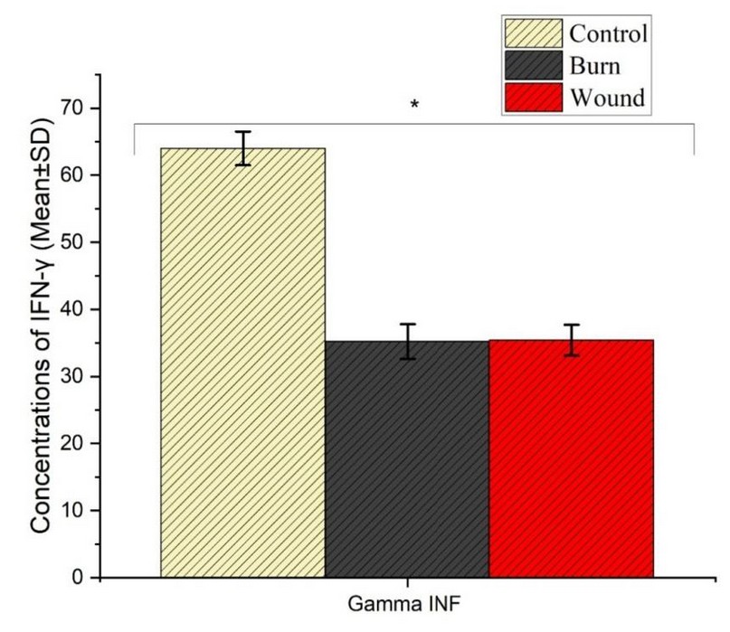
Table 3 show a decrease in the level concentration of Gamma interferon for burn and wound patient with p. aeruginosa infection, where the score mean 35.2±2.6 pg/ml and 35.4±2.3 pg/ml, respectively, when compared with healthy people ( control group) score 46±2.5 pg/ml
The current study agrees with 4 showing the decrease of gamma interferon in a patient infected with Pseudomonas Aeruginosa, and also agrees with 7 when it shows the decrease in interferon – γ level with a patient infected with (PA) compared with no infection; our study also agrees with 8, 9 that reported the decrease in IFN- y in patients with pseudomonas infection. This decrease may result in a suppression of immune system in burn and wound patients afflicted with p.aeruginosa. The immunosuppression includes inhibiting NK cells that produce IFN- y 10; IFN- y are proteins produced by most cells of vertebrates. IFN- y is a critical cytokine for adaptive and innate immunity 4. The current study disagrees with 11. That reported no significant difference between burn patients and the control group.

Table 3. Comparison of Gamma INF means among studied groups (Control, Burn and Wound) * represent a significant difference at p<0.05. letters represent the type of statistical analysis: a; the statistical analysis among all studied groups (Control, Burn and wound), b; the statistical analysis between the Burn group and Control group; c: the statistical analysis between the Wound group and Control group; d: the statistical analysis between Burn group and wound group

Figure 1. Comparison of Gamma INF means among studied groups (Control, Burn and Wound)
CONCLUSIONS
This study showed the decreased production of gamma interferon As a result of a reaction to infection caused by the Pseudomonas aeruginosa in burn and wound patients due to immunosuppression by bacteria study due to virulence factor.
REFERENCES
1. Abd Al-Mayali, M., and Salman, E. D. Bacteriological and Molecular Study of Fluor quinolones Resistance in Pseudomonas aeruginosa isolated from Different Clinical Sources. Iraqi Journal of Science. 2020., 2204-2214
2. Altaai, M. E., Alhadithy, J. A., and Al Swediawy, W. K.. Genetic identification of Opr I and Opr L Genes in Pseudomonas aeruginosa isolated from different local sources. Journal of university of Anbar for Pure science, 2017. 11(1).
3. Aleksiewicz, R., Kostro, K., Kostrzewski, M., Lisiecka, B., Bojarski, M., and Mucha, P. A. Percentage of CD4+, CD8+, and CD25+ T lymphocytes in peripheral blood of pigs in the course of experimental burns and necrectomy. Journal of Veterinary Research.2015, 59(3), 401-410
4. Risan, F. A., Salih, M. K., and Salih, T. A.. Estimation of IL-17A and IFN-y in the Burn of Patients that Afflicted by Different Bacterial Types. International Journal of Psychosocial Rehabilitation. 2020, 24(05).
5. Che Soh, N. A., Rapi, H. S., Mohd Azam, N. S., Santhanam, R. K., Assaw, S., Haron, M. N., ... and Ismail, W. I. W. Acute Wound Healing Potential of Marine Worm, Diopatra claparedii Grube, 1878 Aqueous Extract on Sprague Dawley Rats. Evidence-Based Complementary and Alternative Medicine, 2020
6. McDonald, J.H.. Handbook of Biological Statistics. 3rded. USA: 2014 Sparky House Publishing
7. Al-Jabouri, R., Saqban, A. K. S., Hasson, S. O., and Abady, N. R. IL-18 Act as a Costimulus for Production of Interferon Gamma During Stimulation by. Pseudomonas aeruginosa Infecfion, J Pure Appl Microbiol. 2019,
8. Schnieder, David, F; Cavin, H; Glenn, Douglas, E; and Faunce. Innate Lymphocyte subsets and their Immunoregulatory roles in burn injury and sepsis. Journal of Burn care and Research. 2010
9. Sundararajan, V. Modihally; Mehment Toner; Martin, L. Yarmaush; and Richard, N. Mitchell. Interferon-Gamma modulates trauma induced muscle wasting and immune dysfunction. Nov. 2002, 236(5): 649
10. Couper, KNDG Blount; and Riley, E.M. 2008. IL-10 the master regulator of immunity to infection.
11. Abd-Alrazak. Lina Qays Yaseen. 2017. Bacteriological study of wounds, Burns, and urinary tract
Received: 20 July 2022 / Accepted: 15 October 2022 / Published:15 November 2022
Citation: Jwad M A , Gharbi W A. Determination of IFN-y in Patients with Pseudomonas aeruginosa-Inflicted Burn and Wound. Revis Bionatura 2022;7(4) 2. http://dx.doi.org/10.21931/RB/2022.07.04.2
