2023.08.01.47
Files > Volume 8 > Vol 8 No 1 2023
Detection of lukf-pv gene in Staphylococcus aureus isolated from pregnant women with Urinary tract infection

Hiba Qasim Hameed 1 , Inas Ahmed Saeed2, Enas Abdalhadi Hussain2 *
1 Department of Biology, College of Education for pure science Ibn-Al Haitham, University of Baghdad, Baghdad, Iraq.
2 Department of Microbiology ,Collage of Medicine, University of Babylon
3 Department of Biology, College of Education for pure science Ibn-Al Haitham, University of Baghdad, Baghdad, Iraq.
*Correspondence: [email protected]
Available from: http://dx.doi.org/10.21931/RB/2023.08.01.47
ABSTRACT
Staphylococcus aureus is a human pathogen as well as commensal bacteria. S. aureus has colonized around 30% of the human population. This study aimed to diagnose Staphylococcus aureus by molecular techniques, correlate the resistance against selected antimicrobial substances with the presence of the lukf-pv gene, and find the sequence of lukf-pv gene for the isolates obtained to investigate the mutations of those obtained isolates. This study included 60 patients diagnosed by the hospital with a urinary tract infection in Teaching Medical City Hospital, Baghdad, and Al-Yarmouk Teaching Hospital, between January 2021 and July 2021. The isolates were cultured on a blood agar overnight; then, isolates were diagnosed by VITEK as S. aureus. DNA has been isolated from all the included samples. A specific region of the 16SRNA gene has been amplified to diagnose S. aureus by molecular techniques. Then possession of the lukf-pv gene was tested by PCR, then amplified products were sequenced to detect the mutations within the lukf-pv gene. The finding appeared that blood group O+ has the highest rate of bacterial infection, the lowest is O- (1.7%), and the highest rate is shown within people not suffering from complicated diseases (65%). Of the 60 isolates, 60 (100%) were confirmed by 16sRNA gene amplification and were positive, among which 37 (61.6%) were lukf-pv positive. Results of the lukf-pv gene sequences showed around 501 bits score and 96% compatibility (ID: CP076105.1). The current study showed that antibiotics Cefoxitin, Benzylpenicillin, Oxacillin, Clindamycin, Fusidic acid, Rifampicin, Erythromycin, Vancomycin, and Teicoplanin had the highest resistance to antibiotics and as follow;100 %, 100 %, 40.54 %, 27.03 %, 27.03 %, 16.22 %, 13.51 %, and 10.81 %, respectively.
Keywords: Staphylococcus aureus, 16sRNA and lukf-pv genes
INTRODUCTION
Staphylococcus aureus (S. aureus) is a common human pathogen that causes infections ranging from minor skin and soft-tissue infections to life-threatening sepsis. It has been linked to significant morbidity and mortality in community-acquired (CA) and healthcare-associated (HA) infections1,2.
Surface colonization is a significant risk factor for infection; S. aureus colonization of the skin and mucus membranes is estimated in 30% of healthy people, with another 30% being temporary carriers 3. Because of the widespread use of beta-lactams in infection treatment, S. aureus can acquire methicillin resistance, first in hospitals and, more recently, in community strains 4.
Several cell surface and secreted virulence factors contribute to the pathogenesis of S. aureus. For example, panton-Valentine leukocidin (pvl encodes lukS-PV/lukF-PV genes) is a two-component toxin that causes hole development in complement receptors on leukocyte cell membranes 5. pl-associated S. aureus (pvl-sa) infection is most typically associated with community-acquired methicillin-resistant S. aureus (CA-MRSA) infection 6; however, outbreaks due to pvl-positivity's methicillin-susceptible S. aureus (MSSA) strains have been observed in close community settings 7. This study aimed to identify Staphylococcus aureus by molecular techniques by the 16sRNA gene, then correlate the resistance against selected antimicrobial substances with the presence of the luk gene; this gene has been further investigated by sequencing to investigate the mutations of those obtained isolates.
MATERIALS AND METHODS
Sample Collection
The current research included 60 swabs isolated from pregnant women suffering from UTI attending Teaching Medical City Hospital, Baghdad Teaching Hospital, and Al-Yarmouk Teaching Hospital between January 2021 and July 2021. The bacteria from all 60 nasal swabs were grown on blood agar using the streak way and incubated for 24 hours at 37°C. Standard sterile saline was introduced to the test tube (3.0 mL). An application stick or sterile brush was employed to transfer enough pure-crop colonies and suspend the isolated colonies in normal saline. McFarland's turbidity (0.5-0.63) was adjusted, and Densi ChekTM was employed. The transmission tube is then inserted into the matching suspension tube, while the test tube is placed in the next slot. Suspension cards for bacteria before loading are inoculated by a process that cuts the transfer tube and seals it to the incubator. There is room for up to 60 cards or up to 30 cards. Each card was incubated at 35,5+ 1,0°C for 15 minutes before being removed from the incubator and transferred to the optical system for reaction measurements.
Additionally, a single algorithm is used to remove bogus measures from tiny bubbles. Raw data calculations are performed and compared for each test to evaluate reactions. The test reaction results are displayed on the VITEK 2 Compact as "+," "(-)," or "(+)." Test results of an unknown organism were compared to the associated database to generate a quantitative nearness value for each database taxonomy. Antimicrobial resistance patterns were determined using the VITEK-2 AST.
Detection of 16sRNA and lukf-pv gene by Polymerase Chain Reaction (PCR)
The 16s rRNA and lukf-pv gene were detected using PCR on all S. aureus isolates. For the amplification of 16sRNA gene, a specific primer has been used; the sequence of sense primer, 5′-AACTCTGTTATTAGGGAAGAACA-3′ and the antisense; 5′-CCACCTTCCTCCGG TTTGTCACC-3′ (8). The reaction mixture has been set to 25 µl and contains; Taq PCR PreMix 5µl, Forward primer 10 picomols/µl (1µl), Reverse primer 10 picomols/µl (1µl) DNA 1.5µl (2ng/µl), and Nuclease – Free Water 16.5 µl. The cycling conditions of amplification were as follows; Initial Denaturation at 94ᵒC for 5 min, then 30 cycles of Denaturation -2 at 94ᵒC for 1min, annealing at 55ᵒC for 1min, and extension at 72ᵒC for 2min. Then one cycle of extension at 72ᵒC for 10min. Amplification of lukf-pv gene was done by using the sense primer; 5′- ATCATTAGGTAAAATGTCTGGACATGATCCA-3′, and the antisense primer, 5′-GCATCAAGTGTATTGGATAGCAAAAGC-3′ 8. The reaction mixture is composed of the same components of 16sRNA except for primers. The cycling condition was as follows; Initial Denaturation at 94ᵒC for 3 min. Then 35 cycles of Denaturation at 94ᵒC for 30 seconds, followed by annealing at 55ᵒC for 30 seconds, then extension at 72ᵒC for 30sec. Then one step of extension at 72ᵒC for 7 min. The Macrogen company then sequenced the amplified products.
RESULTS AND DISCUSSION
The results appeared 60 samples isolated from pregnant were diagnosed as S. aureus. It examined the resistance of 37 isolates against antibiotics studies; according to the effects of Viteck, these isolates were resistant to Oxacillin; therefore, S. aureus was considered methicillin-resistant. The percentage of infection with S. aureus has been classified according to the patient's job, and the results are shown (Table 1). The housewife had the highest rate of bacterial illness, 31 (51.7%), followed by 26 Employees (43.3%) and 3 Students (5%), respectively. The difference between the percentage of infection is significant (p<0.0001).
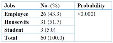
Table 1. The percentage of samples distribution according to jobs.
Antigen expression differences between blood groups can increase or decrease a host's susceptibility to various illnesses. Blood groups can play a direct role in infection by acting as receptors or co-receptors for bacteria, parasites, and viruses. This study showed that blood group O+ has the highest bacterial infection rate, and the lowest is O- (1.7%), as shown in table (Table 2).
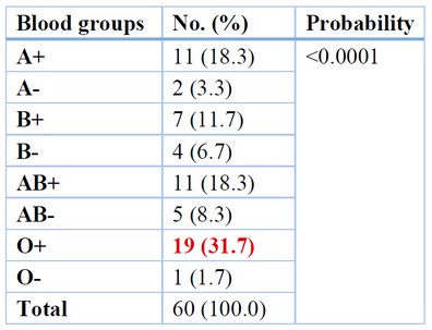
Table 2.Distribution of samples according to Blood groups.
The infection rate differed among the different diseases, as shown in table (3); the highest rate is shown among people that are not suffering from any complicated diseases (65%), followed by people with blood pressure (13.3%), diabetes (11.7) and the lower rate with the Joints infection (10%).
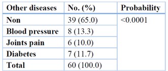
Table 3. Distribution of samples according to diseases.
The electrophoresis results of the amplified product of the 16sRNA region are shown in figure (1). The results showed a clear, sharp band at 756bp for the positive samples, and the results showed the 37 (61.6%) S. aureus isolates under study have the gene lukf-pv from all 60 isolates. In contrast, the results of electrophoresis of the amplified lukf-pv gene showed 433bp in Figure 2.
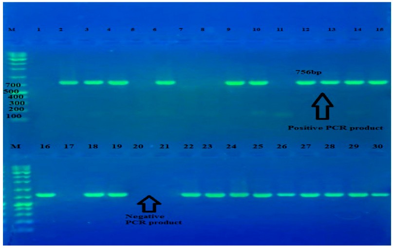
Figure 1. The PCR product (756bp) of 16sRNA gene was electrophoresis on 2% agarose at 5 volt/cm2. 1x TBE buffer for 1:30 hours. N: DNA ladder (1000bp).
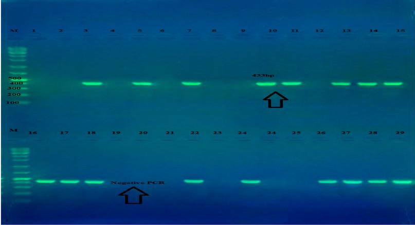
Figure 2. The PCR product (433bp) of lukf-pv gene was electrophoresis on 2% agarose at 5 volt/cm2. 1x TBE buffer for 1:30 hours. N: DNA ladder (1000bp).
To demonstrate the resistance of bacterial strains against the antibiotic, the antibacterial sensitivity of S. aureus isolates for 19 antibiotics was tested using the Vitek- AST methods, and the results of the current investigation revealed that some S. aureus isolates had a clear difference in the antibiotic resistance utilized in this study (Table 4). It was observed from the results of the present study that the highest resistance to antibiotics was to the antibiotics Cefoxitin, Benzylpenicillin, Oxacillin, Clindamycin, Fusidic acid, Rifampicin, Erythromycin, Vancomycin and Teicoplanin; 100%,100%, 100%, 40.54%, 27.03%, 27.03%, 16.22%,13.51%, 10.81% respectively.
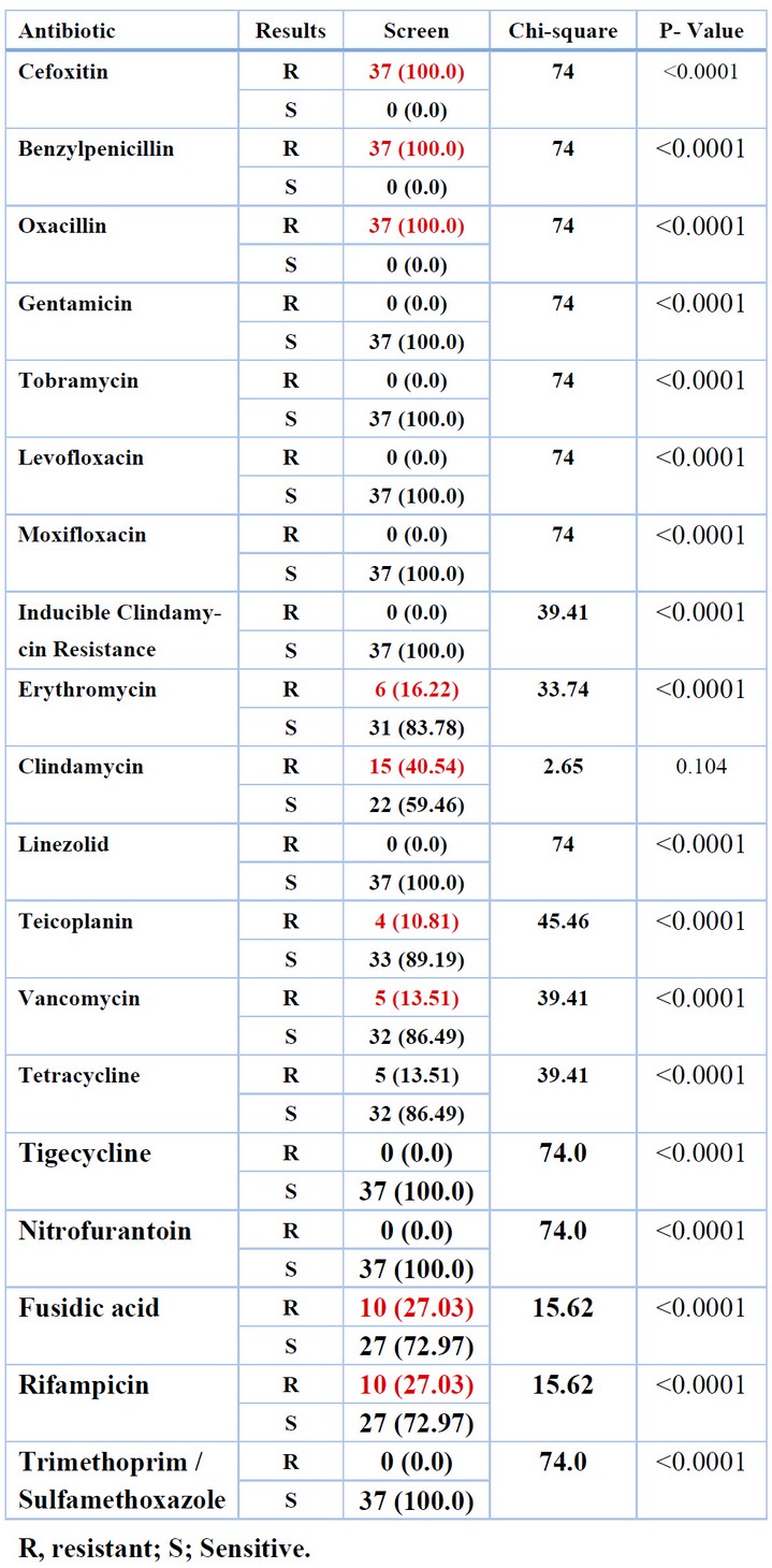
Table 4. The percentages of antibiotic resistance of S. aureus isolates
luk-pvl gene sequences around 501 bits sore showed 96% compatibility (ID: CP076105.1), as seen in figure (3), while varied in 1%. And this percentage of difference includes 14 variations; two Transition A>G, the first one is 81bp , and the second transition is 105bp.Two Transversion share the same changing code, which is T>A and located in (49, 76 nucleotide), and Three Transversions T>G in locations (51, 77,110 nucleotide) , as well as one Transversion G>C in location (45 nucleotide), also three deletion C\-,A\-, C\- in locations (24,40,44nucleotide), and one Insertion -\G in location (9nucleotide).
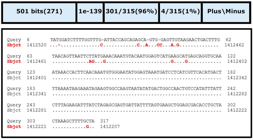
Figure 3. Alignment analysis luk-pv-1 gene of S. aureus. Query symbolizes the gene sequence of the sample; Subject represents the gene sequence by the database of NCBI.
In the present study, the results showed for S. aureus carriers, 61.6% of the isolates were identified as MRSA that carried the Luk-pv-1 gene, disagreeing with Hussein et al. 9 the results showed 22.5% in Iraq's Kurdistan Region; this agreement with findings of a prior study conducted in Duhok in 2016 10.
In Iran, the prevalence of MRSA was 16 - 35% (6,7). The MRSA rate was 10.1% in Jordan 11, and 73% in Saudi Arabia 12; in another study in Iraq that was conducted in rural and urban schools, the rates of nasal infection by isolates of S. aureus and MRSA were 17.75% and 10%, respectively 13.
MRSA strains obtained from hospitals in the Netherlands had a pvl positive 8% of the isolates carrying the pvl gene. In Iran, MRSA, the pvl gene, and MRSA-pvl isolates were found in 19-32, and10 %, of the population, respectively 14. In this study pvl prevalence (61.6%) was extremely high, but similar to that reported in Ghana (75 %) 15, and higher than that recorded in Senegal (47 %), elsewhere in Africa, and around the world 1,3. According to research by Akanbi et al. 16 Antibiotic resistance in S. aureus varied greatly, with the highest rates of resistance to rifampicin and clindamycin (80 %), and erythromycin (70 percent) disagree with our findings, whereas Oxacillin (73.3%) and cefoxitin (76.7%) this agree with our study. Multiple antibiotic-resistant patterns were found in all 37 S. aureus isolates (resistant to three or more antibiotics). In a survey conducted by Velasco et al., 8a nasal infections rate of 7.6% for S. aureus was detected, which is significantly lower than the prevalence reported, contradicting our findings agree with those of other studies 17,18 S. aureus bacteria are resistant to three or more antibiotics.
CONCLUSIONS
It was concluded that Staphylococcus aureus isolated from pregnant with urinary infections were resistant to antibiotics studied. Thirty-seven isolates were resistant against antibiotics studies and resistant to Oxacillin; therefore, S. aureus was considered methicillin-resistant. Moreover, S. aureus isolates have the gene lukf-pv.
Funding: No
Conflicts of Interest: There are no conflicts
REFERENCES
1. Breurec S, Fall C, Pouillot R, Boisier P, Brisse S, Diene-Sarr F, Djibo S, Etienne J, Fonkoua MC, Perrier-Gros-Claude JD, Ramarokoto CE. Epidemiology of methicillin-susceptible Staphylococcus aureus lineages in five major African towns: high prevalence of Panton-Valentine leukocidin genes. Clinical Microbiology and Infection. 2011 Apr 1;17(4):633-9.
2. Özekinci T, Dal T, Yanık K, Özcan N, Can Ş, Tekin A, Yıldırım Hİ, Kandemir İ. Panton-Valentine leukocidin in community and hospital-acquired Staphylococcus aureus strains. Biotechnology & Biotechnological Equipment. 2014 Nov 2;28(6):1089-94.
3. Shallcross LJ, Fragaszy E, Johnson AM, Hayward AC. The role of the Panton-Valentine leucocidin toxin in staphylococcal disease: a systematic review and meta-analysis. The Lancet infectious diseases. 2013 Jan 1;13(1):43-54.
4. Cosgrove SE, Sakoulas G, Perencevich EN, Schwaber MJ, Karchmer AW, Carmeli Y. Comparison of mortality associated with methicillin-resistant and methicillin-susceptible Staphylococcus aureus bacteremia: a meta-analysis. Clinical infectious diseases. 2003 Jan 1;36(1):53-9.
5. Rasigade JP, Laurent F, Lina G, Meugnier H, Bes M, Vandenesch F, Etienne J, Tristan A. Global distribution and evolution of Panton-Valentine leukocidin-positive methicillin-susceptible Staphylococcus aureus, 1981–2007. The Journal of infectious diseases. 2010 May 15;201(10):1589-97.
6. Asiimwe BB, Baldan R, Trovato A, Cirillo DM. Molecular epidemiology of Panton-Valentine Leukocidin-positive community-acquired methicillin resistant Staphylococcus aureus isolates in pastoral communities of rural south western Uganda. BMC infectious diseases. 2017 Dec;17(1):1-7.
7. Rasigade JP, Laurent F, Lina G, Meugnier H, Bes M, Vandenesch F, Etienne J, Tristan A. Global distribution and evolution of Panton-Valentine leukocidin-positive methicillin-susceptible Staphylococcus aureus, 1981–2007. The Journal of infectious diseases. 2010 May 15;201(10):1589-97.
8. Velasco V, Buyukcangaz E, Sherwood JS, Stepan RM, Koslofsky RJ, Logue CM. Characterization of Staphylococcus aureus from humans and a comparison with isolates of animal origin, in North Dakota, United States. PloS one. 2015 Oct 20;10(10):e0140497.
9. Hussein N, Salih RS, Rasheed NA. Prevalence of methicillin-resistant Staphylococcus aureus in hospitals and community in Duhok, Kurdistan region of Iraq. International Journal of Infection. 2019 Apr 30;6(2).
10. Hussein NR. Prevalent genotypes of Staphylococcus aureus strains isolated from healthcare workers in Duhok City, Kurdistan Region, Iraq. International Journal of Infection. 2016 Apr 1;3(2).
11. Aqel AA, Alzoubi HM, Vickers A, Pichon B, Kearns AM. Molecular epidemiology of nasal isolates of methicillin-resistant Staphylococcus aureus from Jordan. Journal of Infection and Public Health. 2015 Jan 1;8(1):90-7.
12. Iyer A, Kumosani T, Azhar E, Barbour E, Harakeh S. High incidence rate of methicillin-resistant Staphylococcus aureus (MRSA) among healthcare workers in Saudi Arabia. The Journal of Infection in Developing Countries. 2014 Mar 13;8(03):372-8.
13. Hussein NR, Basharat Z, Muhammed AH, Al-Dabbagh SA. Comparative evaluation of MRSA nasal colonization epidemiology in the urban and rural secondary school community of Kurdistan, Iraq. PLoS One. 2015 May 1;10(5):e0124920.
14. Lari AR, Pourmand MR, Ohadian Moghadam S, Abdossamadi Z, Namvar AE, Asghari B. Prevalence of PVL-containing MRSA isolates among hospital staff nasal carriers. Laboratory Medicine. 2011 May 1;42(5):283-6.
15. Dekker D, Wolters M, Mertens E, Boahen KG, Krumkamp R, Eibach D, Schwarz NG, Adu-Sarkodie Y, Rohde H, Christner M, Marks F. Antibiotic resistance and clonal diversity of invasive Staphylococcus aureus in the rural Ashanti Region, Ghana. BMC Infectious Diseases. 2016 Dec;16(1):1-6.
16. Akanbi OE, Njom HA, Fri J, Otigbu AC, Clarke AM. Antimicrobial susceptibility of Staphylococcus aureus isolated from recreational waters and beach sand in Eastern Cape Province of South Africa. International journal of environmental research and public health. 2017 Sep;14(9):1001.
17. Gorwitz RJ, Kruszon-Moran D, McAllister SK, McQuillan G, McDougal LK, Fosheim GE, Jensen BJ, Killgore G, Tenover FC, Kuehnert MJ. Changes in the prevalence of nasal colonization with Staphylococcus aureus in the United States, 2001–2004. The Journal of infectious diseases. 2008 May 1;197(9):1226-34.
18. Mainous AG, Hueston WJ, Everett CJ, Diaz VA. Nasal carriage of Staphylococcus aureus and methicillin-resistant S aureus in the United States, 2001–2002. The Annals of Family Medicine. 2006 Mar 1;4(2):132-7.
Received: December 23, 2022 / Accepted: January 30, 2023 / Published:15 February 2023
Citation: Hameed H Q, Saeed I A, Hussain E A. Detection of lukf-pv gene in Staphylococcus aureus isolated from pregnant women with Urinary tract infection. Revis Bionatura 2023;8 (1)47. http://dx.doi.org/10.21931/RB/2023.08.01.47