2022.07.04.57
Files > Volume 7 > Vol 7 No 4 2022
Efficacy of zinc oxide nanoparticles and Bifidobacterium bifidum Extraction on anaerobic bacteria isolated from patients with diarrhea
1,4Department of Biology, College of Science, Tikrit University, Salah al-Din –Iraq
2Department of Biology, College of Science, Mosul University, Ninive- Iraq
3Ministry of Health, Salah al-Din General Hospital, Salah al-Din –Iraq
*Corresponding author: [email protected],[email protected],
Available from: http://dx.doi.org/10.21931/RB/2022.07.04.57
ABSTRACT
Infectious acute diarrhea may be prevented with probiotics; because they make up the majority of the colonic flora in breastfed newborns and are likely to contribute to the lower incidence of diarrhea in this population, Bifidobacteria are particularly appealing as probiotics agents. The present study was designed to identify anaerobic bacteria, especially C. difficile the main reason for dysentery associated with antibiotics. Detect the ability of each ZnoNPs and B. bifidum to inhibit bacterial growth. During the period from March to October 2019, (100) children and adults who came to Salah al-deen hospital in Tikrit city participated in the study under the supervision of a physician. All samples were transported using a carry Blair if late one or two hours after collection and culturing. The collected models were also cultured on Xylose Lysine Deoxycholate agar, Salmonella Shigella agar, Eosin methylene blue agar, and MacConkey agar. For initiated diagnoses of the Enterobacteriaceae, Blood agar is used to detect beta-hemolytic isolates, recover enteric bacteria other than Enterobacteriaceae, and evaluate the results of oxidase tests. To diagnose bacterium kinds, biochemical reactions and motility tests were used. Impact of ZnoNPs, and B. bifidum antibiotic In vitro. The results of 100 dysentery feces samples were obtained into (60%) samples for males and (40%) for females. Eighty-two positively impacted anaerobically on growth media like Clostridium complicate agar and MacConkey agar (18%) other than bacteria.
In contrast, negative samples revealed 10 (55.56 percent) samples for males and 8 (44.44 percent) samples for females. The same stool samples were taken and cultured on Clostridium difficile agar and MacConkey agar under anaerobic and ideal incubation conditions. 15% and 67% of isolates appeared on MacConkey agar of the total number of samples, while 18% showed negative growth. Finally, Zn NPs showed their ability to inhibit Clostridium complicated segregate lean on the condensation 5 mg/ml, and it caused the inhibitory effect on Clostridium to complicate by 10-22 of the diameter of inhibition. The Inhibition Zone Dimeter ranged from 8 to 25 mm for isolates when condensation was utilized at 2.5 mg/ml. According to the findings, the widths of the inhibitory zones for isolates of C. difficile containing B. bifidum supernatant mg/ml ranged from 9 to 24 mm.
Keywords: Zinc oxide nanoparticles, Probiotic, Bifidobacterium bifidum, Clostridium difficile
INTRODUCTION
Diarrhea is a confusing matter in older people, especially in people with general weakness due to other problems in the body, such as stool or fluid electrolyte disturbances. A complication associated with the use of antibiotics is Clostridium difficile infection (CDI)1. Intestinal flora dominates. Using broad-spectrum antimicrobials may destroy the patient's normal flora and encourage the dissemination of C. difficile toxins. Thus, antimicrobial therapy is critical to the development of CDI 1. In the last year, many studies using Nanotechnology as antimicrobial activity and zinc oxide nanoparticles as an antibacterial factor appeared in biologists' medicine, chemists and physicists 2. Scientists have searched for many years for antibacterial agents for Helicobacter pylori, for examples 3, 4. A significant reduction in apoptosis was demonstrated in infants with viscera 5. Changes to the body's defensive 6 , 7 Measures to reduce inflammation reduced visceral-induced morbidity, as well 8 ,9. Hence, this study identified the types of aerobic bacteria that cause diarrhea. Detect the ability of each ZnoNPs and B. bifidum to inhibit bacterial growth. The current investigation aimed to identify anaerobic bacteria, particularly C. difficile, the primary cause of diarrhea linked with antibiotic use. Find out whether ZnoNPs and B. bifidum can both stop bacterial growth.
MATERIALS AND METHODS
Sample collection
Samples were taken from patients of different age groups in Salah al-Din Hospital in Tikrit governorate from March 2019 to October 2019; the study included 100 samples, and vectors were used for transplanting them on the following media 10. For initial isolation of Enterobacteriaceae, such as MacConkey agar; XLD agar, SS agar, and EMB agar; and blood agar to disclose beta-hemolytic isolates recover enteric bacteria other than Enterobacteriaceae, as well as oxidase test performance10. Aerobic and anaerobic isolates were incubated in a jar at 37 C° for 24 hours, and all biochemical tests were performed to detect Gram-positive and negative species, as well as a motility test 11.
ZnO NPs and Bifidobacterium bifidium were shown to have a minimal inhibitory concentration.
ZnO NPs and Bifidobacterium bifidius were studied according to the agar Dilution method. The concentrations of ZnO in the supernatant of Bifidobacterium bifidum varied from 1-2 mg/ml. Muller Hinton agar center in glass bottles (20 ml each), sterilized by inventory and returned to a temperature of 4 Co. Different amounts of ZnO and Bifidobacterium bifidus were added, the media was fed correctly, and then the media was placed on sterile plates and maintained at 4 C° until use. The infection was caused by bacteria examined and compared to a set turbidity standard solution after 18-24 hours. The micropipette of five μl was made from the micropipette, then the dishes with various dosages of ZnO and Bifidobacterium bifidus were extracted, and the dishes were allowed for a while before drying. Dishes were incubated upside down at 37°C for 24 hours. After incubation, determine the minimum inhibitory concentration (MIC), the lowest concentration of the chemical utilized that did not result in noticeable bacterial growth or the development of only a small number of bacterial colonies 12.
In vitro assessment of the efficacy of ZnoNPs and Bifidobacterium bifidus as microbial antibiotics
Fifteen isolates were selected from different stool samples and tested for the effectiveness of ZnONPs on them, as reported in NCCLS (2004), which included: Preparation of bacterial suspension from samples treated with the treatment (Bifidibactrium bifidum and ZnO). It was compared with the tubes containing McFarland standard (0.5)13 to obtain a dilution of 1.5x 10 8 colony forming units (CFU/ml) and then taken from it (0.1 ml) and planted on (Muller Hinton agar) and distributed on the surface of the medium and left for 15 minutes. Then, 100 μl of each treatment were added to ZnO NPs and Bifidobacterium bifidum at a concentration of 2 mg/ml, and the plates were incubated at 37 °C for 24 hours. Next, the inhibition zone was evaluated14.
Statistical Analysis
The SPSS 15, IBM version 20, program was used to perform the analysis on the data18. Statistical significance is assumed when the p-values are less than 0.05.
RESULTS
Sample distribution
Table (1) shows the growth of anaerobic species from stool samples in both gender; 100 stool samples were collected and distributed among 60 males and 40 females. Anaerobic samples showed positive growth of gram stain on each of Clostridium difficile agar and MacConkey agar medium. At the same time, 18% of the samples showed a non-bacterial increase, distributed among (55.56%) samples for males and 8 (44.44%) for females.

Table 1. The growth of anaerobic species from stool samples in both gender
Bacterial Isolation
Under anaerobic conditions at 37°C, a hundred patient samples were grown on Clostridium difficile agar and MacConkey agar. It causes bacterial isolates to emerge on 15 % and 67 % of the total samples on MacConkey agar, respectively, whereas 18 % showed no growth on any of the utilized mediums.
Diagnoses of Clostridium difficile.
The bacterial isolates appear flat and have a ground-glass appearance, which is apparent on the Clostridium difficile agar and TCCFA medium. According to the results of bacterial transplanting, the bacteria manifested as circular, convex colonies with distinct borders and a gray tint. The bacteria were positive bacilli according to the results of the pigmentation gram stain. The results of biochemical identification, such as catalase and oxidase, were found to be negative, whereas gelatin liquefaction was determined to be positive (gelatin liquefaction +ve). In addition, the fermentative of glucose, fructose, and mannose changed color, indicating positive findings, and the enzymatic reactions were negative for casein hydrolysis. Still, positive results for lecithinase and lipase were detected for the esculin hydrolysis test, as shown in table (2).
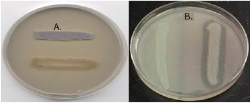
Figure 1: A. Lipase test , B. lecithinase test

Table 2. Biochemical tests for Clostridium difficile
Diagnoses of Enterobacteriaceae species.
The shape and diameter indicate bacterial isolates grown anaerobically on MacConkey agar for 18-24 h (Figure. 2) to the following Enterobacteriaceae. The diagnosis was based on microscopic examination using Gram stain. The appearance of Gram-negative species, which are given to these species as references to Enterobacteriaceae. The production of the capsule and characteristics of the colony, such as mucus and metallic luster, all these parameters were used to determine the bacterial genus. The hemolysis, catalase, oxidase, urease production, and IMViC test were performed, according to 11. The study showed that 100 samples of cultured stool and 67 samples gave a positive culture with enteric isolates bacteria that caused diarrhea in patients. The triple sugar iron agar test uses sugar and iron to diagnose different types of intestinal bacteria. Containing three fermentable sugars, the isolates were diagnosed by the appearance of the characterization results. Escherichia coli was a response to Triple Sugar Iron agar. While Pseudomonas and Shigella Sonni can't ferment it, the results are Alk/Alk, Alk/Alk, with no gas and no H2S generation. K.pneumoniea was also found to alter medium to Acid/Acid, produce gas, although it was variable (d) to produce H2S. While Serriatia marcessens appeared to be Alk/Acid, produced gas, and was unable to create H2S. These bacteria exploited the egg yolk suspension in the medium to produce lecithinase and lipase, which were also employed in diagnostics.
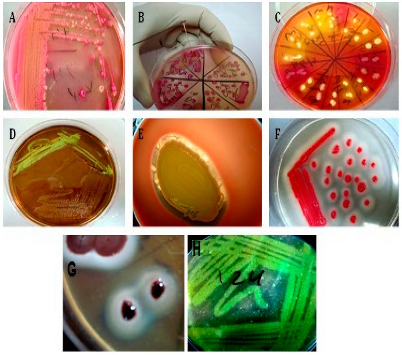
A-Pink colonies represent lactose fermenter, while pale colonies represent non-lactose fermenter colonies on differential and selective Mac Conkey agar media.
B- Mucoid characters of Klebsiella spp. on Mac Conkey agar media.
C- Organisms such as E. coli and Klebsiella-Enterobacter species may utilize more than one carbohydrate and produce bright yellow colonies, species that use non of the carbohydrate produce translucent colonies. Most species of Salmonella have red colonies, most with black centers from H2S gas and Citrobacter colonies are yellow with black centers.
D- Typical strong lactose fermenter, notably E. coli, produce colonies that are green-black with a metallic sheen on Eosin methylene blue agar (EMB)
E-The clear zone around the swarming colony represents beta-hemolysis on blood agar.
F-The opacity around the squeaking culture represents lipase producer Serratia marsessence on Sierra media.
G-The opacity around the colony represents lecithinase producer bacteria on Egg yolk agar media.
H- The clear zone on Skim milk agar around the streaking culture represents protease-producing bacteria.
Figure 2. Primary, selective and differential media are used for bacterial isolation and identification.
C. defficile isolated from patients with diarrhea was inhibited by ZnoNPs
Table (3) and figure 3 demonstrate the inhibitory effects of zinc oxide particles (ZnO NPs) on C. difficile isolates from individuals with diarrhea (5). The metronidazole antibiotic was chosen for its distinct mode of action against C.difficile isolated from diarrhea patients, and the outcomes of the ZnO NPs inhibition investigation were contrasted with those of this drug. Additionally, the diameter of the inhibitory zone served as the basis for the measurement (IZD). The results demonstrated that, depending on the concentration, ZnO NPs could inhibit C.difficile isolates, with a concentration of 5 mg/ml inhibiting C.difficile isolates with diameters ranging from 10 to 22 mm. For the isolates with a concentration of 2.5 mg/ml, the IZD appeared between 8 and 25 mm. Furthermore, only three isolates with an IZD of 5,10,10 mm seemed susceptible to ZnONPs at a concentration of 1.25. While the 0.625 mg/ml concentration of ZnONPs was ineffective in suppressing C.difficile isolates.
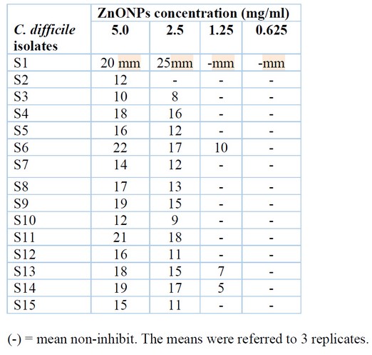
Table 3. Inhibition zone diameter in mm of ZnONPs against C.difficile isolates.
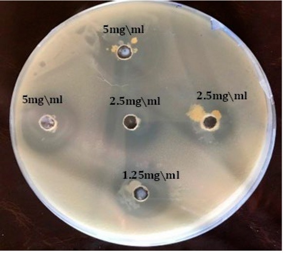
Figure 3. Zno NPs gradient concentration inhibition growth bacteria C. difficile
The C. defficile isolate from diarrhea patients that Bifidobacterium bifidum supernatant inhibits:
The effect of Bifidobacterium bifidum supernatant on the growth of C. difficille isolates from diarrhea patients is shown in Table (4). The inhibition zone diameters IZD of B. bifidum supernatant were observed to be between 9 and 24 mm against C.difficile.
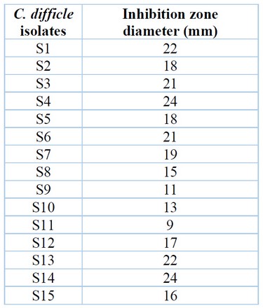
Table 4. The size of the B.bifidus supernatant's inhibitory zone against C.difficle isolates.
DISCUSSION
There are several reasons, including the growth of aerobic bacteria or diarrhea resulting from antibiotics. Despite the tremendous development in diagnostic techniques, many of the causes leading to gastroenteritis have not been explained to this day 16. The other samples from diarrhea patients were positive for bacterial growth in 82 % of cases. Males had 50 positive samples (60.%), while females had 32 (39.02 %), and the male-to-female ratio was 1.2: 1. These findings were confirmed by 17, who discovered that males had 62.26 % diarrhea while females had 37.74 %. It was also agreed with 17, who discovered that 62 percent of males and 38 % of girls in hospitals had diarrhea. Clostridium difficile isolation from diarrhea is extremely rare, owing to the need for a persistent anaerobic environment to promote optimum development. Because it prevents the growth of normal fecal flora, they used the taurocholate-cefoxitin-cycloserine-fructose agar (TCCFA), which is selective and differential for C. difficile 18. By having both cefoxitin, which more broadly prevents the growth of Gram-negative and -positive bacteria, and c. difficile and the majority of enterococci strains, it can act as a bacteriostat for Gram-negative bacteria.
However, 67 % of the bacterial isolates appeared on MacConkey agar media and appeared in various sizes and shapes, indicating that they belonged to the Enterobacteriaceae family of bacteria. There are methods for creating and counting spore stocks in vitro for various downstream applications, including microscopy. Since fructose fermentation lowers pH and changes the medium's color from red/orange to yellow, the pH indicator neutral red can be added 18. The fermentative results of sugars such as glucose, fructose, and mannose led to changed color and were considered positive findings; these results, which show on the parameters above, were conformity for the isolates as C.diffcille 19 The breakdown of the lecithin in the egg yolk causes an opaque precipitate to develop surrounding the colonies. The Lipase enzyme hydrolyzes the lipids in the egg yolk, giving the colony's surface an iridescent shine12. Except for P.flourscence, all isolates appear to be negative for this test. The gelatin hydrolysis test was also done, and all isolates were determined to be negative, except for Serratia marscence, which was found to be positive 20.
Interest in probiotics' potential to control RV diarrhea has grown over the past decades. There is evidence that certain Bifidobacterium strains can treat gastroenteritis in this situation. 21 , 22. However, B. bifidum and other Bifidobacterium strains have effectively reduced viral clearance in babies, 23 even in mice 24. In vitro studies have shown that Bifidobacteria and Lactobacillus can inhibit RV infection, for example, by interfering in the adhesion step. On the other hand, the reduction of viral shedding exerted by Lactobacillus is consistent with other studies, which found that L. rhamnosus GG could reduce RV elimination in gnotobiotic piglets infected with human RV and in children. 25. There was no previous study, at least locally, that demonstrated the ability of ZnONPs to inhibit C.difficile. Still, the ability of ZnONPs to inhibit C.difficile isolated from diarrhea patients is visible, confirming the ability of these nanoparticles were shown to penetrate the cell walls of bacterial isolates, destroying cellular activity and causing cell death. The antibacterial activities of Zn NPs were investigated by determining the lowest inhibitory concentration against E.coli bacteria, which was corroborated by 26. Diffusion in the pits was used to monitor the antibacterial nanoparticle's quantitative assessment, and it was shown that the area of inhibition mostly relied on the concentration and agreement with results by the 14. It showed that bacterial inhibition increased as ZnO NP concentration was raised. The results were also in agreement with 27. The MIC was measured using a concentration of ZnONPs of 1.25 mg/ml, and it was discovered that the MIC against C.difficile isolates was 1.25 mg/ml. While this was discovered, the MBC for the identical bacterial isolates was 2.5 mg/ml. The capacity of ZnO NPs to disrupt the bacterial membrane, resulting in cytosolic component leakage and bacterial cell death, is one of the mechanisms of bacterial inhibition. The findings were in line with those of 28; in dosages of 1.2-1.6 mg/ml, ZnO NPs were shown to be more effective at inhibiting V.cholerae and Enterotoxigenic Escherichia coli (ETEC) 29 discovered that Zn NPs had anticoccidial and antioxidant activities in the jejunum after infection with Eimeria papillata. Gram-negative (Escherichia coli) and Gram-positive (Staphylococcus epidermis) bacteria were shown to have different antibacterial effects from nanoparticles, with Gram-negative bacteria having stronger antimicrobial activity than Gram-positive bacteria 30. The findings of The inhibitory activity of Bifidobacterium bifidum supernatant against C. defficile isolated from diarrhea patients concurred with 31 who showed that probiotics reduce the risk by 42% compared to advanced diarrhea associated with antibiotics. Thus 32 who used a probiotic B. bifidusm to treat mice infected with H. pylori and who used probiotics in the treatment of Clostridium difficile infection agreed that probiotics were beneficial for both adults and children, reducing the risk of diarrhea associated with Clostridium difficile by 59.5 and 59.5. 65.9%, respectively. Several variables contribute to the effectiveness of Bifidobacterium species. Major Probiotic mechanisms of action include competitive exclusion of pathogenic microorganisms, production of anti-microorganism substances, and immune system modulation. They also include epithelial barrier enhancement, increased adhesion to the intestinal mucosa, and concurrent inhibition of pathogen adhesion 33 , 34.
In the previous study, there has been some debate about the possibility of using a wide variety of chemicals to stop the growth of hazardous microbes. One example of this would be the direct effect that Ag and TiO2 nanoparticles had on dangerous bacteria in the earlier study number 35. This would be an illustration of this. The testing of these nanoparticles included the utilization of P. mirabilis and P. vulgaris, examples of bacteria that can be dangerous to humans. On the other hand, several studies have shown that treating harmful bacteria with therapy that involves physical forces, such as audible noises and magnetic fields, can help diminish the resistance of S. aureus to infection 36. Several different researchers carried out these studies. A wide range of researchers from a variety of institutions carried out these investigations. Due to the researchers' efforts, this discovery was made.
CONCLUSIONS
When C. difficile was grown in isolation from diarrhea, our findings led us to the conclusion that zinc oxide nanoparticles and probiotics both have a good effect in preventing the growth of the pathogen. This assumption was proven beyond a reasonable doubt by the use of an approach known as good diffusion in the pits. It was discovered in the lab that increasing the concentration leads to a bigger increase in the total quantity of inhibition that can be seen on the plate. This was one of the discoveries made. Both the bacteria that are responsible for diarrhea and the bacteria that are isolated from diarrhea are candidates for therapy with a variety of enhancers and nanomaterial, according to the research group that conducted the study.
Acknowledgment: We extend our thanks, appreciation and gratitude to everyone who helped write, prepare and publish this research paper and proofread it.
Conflict between authors: No conflict
Funds: self by authors
REFERENCE
1. Pituch KA, Green VA, Didden R, Lang R, O'Reilly MF, Lancioni GE, Sigafoos J. Parent reported treatment priorities for children with autism spectrum disorders. Research in Autism Spectrum Disorders. 2011 Jan 1;5(1):135-43.
2. Suchea M, Christoulakis S, Moschovis K, Katsarakis N, Kiriakidis G. ZnO transparent thin films for gas sensor applications. Thin solid films. 2006 Oct 25;515(2):551-4.
3. Shirasawa Y, Shibahara-Sone H, Iino T, Ishikawa F. Bifidobacterium bifidum BF-1 suppresses Helicobacter pylori-induced genes in human epithelial cells. Journal of dairy science. 2010 Oct 1;93(10):4526-34.
4. Chenoll E, Casinos B, Bataller E, Astals P, Echevarría J, Iglesias JR, Balbarie P, Ramón D, Genovés S. Novel probiotic Bifidobacterium bifidum CECT 7366 strain active against the pathogenic bacterium Helicobacter pylori. Applied and environmental microbiology. 2011 Feb 15;77(4):1335-43.
5. Khailova L, Mount Patrick SK, Arganbright KM, Halpern MD, Kinouchi T, Dvorak B. Bifidobacterium bifidum reduces apoptosis in the intestinal epithelium in necrotizing enterocolitis. American Journal of Physiology-Gastrointestinal and Liver Physiology. 2010 Nov;299(5):G1118-27.
6. Fu YR, Yi ZJ, Pei JL, Guan S. Effects of Bifidobacterium bifidum on adaptive immune senescence in aging mice. Microbiology and immunology. 2010 Oct;54(10):578-83.
7. Philippe D, Heupel E, Blum-Sperisen S, Riedel CU. Treatment with Bifidobacterium bifidum 17 partially protects mice from Th1-driven inflammation in a chemically induced model of colitis. International journal of food microbiology. 2011 Sep 1;149(1):45-9.
8. Mouni F, Aissi E, Hernandez J, Gorocica P, Bouquelet S, Zenteno E, Lascurain R, Garfias Y. Effect of Bifidobacterium bifidum DSM 20082 cytoplasmic fraction on human immune cells. Immunological Investigations. 2009 Jan 1;38(1):104-15.
9. Guglielmetti S, Mora D, Gschwender M, Popp K. Randomised clinical trial: Bifidobacterium bifidum MIMBb75 significantly alleviates irritable bowel syndrome and improves quality of life––a double‐blind, placebo‐controlled study. Alimentary pharmacology & therapeutics. 2011 May;33(10):1123-32.
10. Baron EJ, Peterson R, Finegold SM. Hospital epidemiology, conventional and rapid microbiology methods for identification of bacteria and fungi. Bailey and Scott's Diagnostic Microbiology, Mosby,. 1994:97-108.
11. Jawetz, E., Melnik, J.L. ; Adelberg , E.A.; Brook, G.F.; Butel, J.S. and Morse , S.A. (2010). Medical Microbiology 26th. ed. Appleten and Lang New York. Connctical. PP.45-60 .
12. Wu W, Huang W, Wei A, Schmidt M, Krause U, Wu D. Inhibition effect of N2/CO2 blends on the minimum explosion concentration of agriculture and coal dusts. Powder Technology. 2022 February 1;399:117195.
13. MacFaddin JF. Biochemical tests for identification of medical bacteria, 3rd ed. Lippincott Williams and Wilkins, USA. Philadelphia, PA. 2000;113.
14. Hamad AM, Atiyea QM. In vitro study of the effect of zinc oxide nanoparticles on Streptococcus mutans isolated from human dental caries. In Journal of Physics: Conference Series 2021 May 1 (Vol. 1879, No. 2, p. 022041). IOP Publishing.
15. Statistics IS. IBM Corp. Released 2020. IBM SPSS Statistics for Windows, Version 22.0. Armonk, NY: IBM Corp. Armonk, NY: IBM Corp.
16. Pimentel M, Chow EJ, Lin HC. Normalization of lactulose breath testing correlates with symptom improvement in irritable bowel syndrome: a double-blind, randomized, placebo-controlled study. The American journal of gastroenterology. 2003 Feb 1;98(2):412-9.
17. Alrifai SB, Alsaadi A, Mahmood YA, Ali AA, Al Kaisi LA. Prevalence and etiology of nosocomial diarrhoea in children 5 years in Tikrit teaching hospital. EMHJ-Eastern Mediterranean Health Journal, 15 (5), 1111-1118, 2009. 2009.
18. Atlas, MR Handbook of Media for Invironmental Microbiology.2nd ed .Taylor & Francis publisher.New York. USA, 2005, 157-189.
19. Eberly AR, Elvert JL, Schuetz AN. Best Practices for the Pre-Analytic Phase of Anaerobic Bacteriology. Clinical Microbiology Newsletter. 2022 Apr 1;44(7):63-71.
20. George P, Gupta A, Gopal M, Thomas L, Thomas GV. Multifarious beneficial traits and plant growth promoting potential of Serratia marcescens KiSII and Enterobacter sp. RNF 267 isolated from the rhizosphere of coconut palms (Cocos nucifera L.). World Journal of Microbiology and Biotechnology. 2013 Jan;29(1):109-17.
21. Gonzalez-Ochoa G, Flores-Mendoza LK, Icedo-Garcia R, Gomez-Flores R, Tamez-Guerra P. Modulation of rotavirus severe gastroenteritis by the combination of probiotics and prebiotics. Archives of microbiology. 2017 Sep;199(7):953-61.
22. Rigo-Adrover M, Saldaña-Ruíz S, Van Limpt K, Knipping K, Garssen J, Knol J, Franch A, Castell M, Pérez-Cano FJ. A combination of scGOS/lcFOS with Bifidobacterium breve M-16V protects suckling rats from rotavirus gastroenteritis. European journal of nutrition. 2017 Jun;56(4):1657-70.
23. Saavedra JM, Bauman NA, Perman JA, Yolken RH, Oung I. Feeding of Bifidobacterium bifidum and Streptococcus thermophilus to infants in hospital for prevention of diarrhoea and shedding of rotavirus. The lancet. 1994 Oct 15;344(8929):1046-9.
24. Duffy LC, Zielezny MA, Riepenhoff-Talty M, Dryja D, Sayahtaheri-Altaie S, Griffiths E, Ruffin D, Barrett H, Rossman J, Ogra PL. Effectiveness of Bifidobacterium bifidum in mediating the clinical course of murine rotavirus diarrhea. Pediatric Research. 1994 Jun;35(6):690-5.
25. Azagra-Boronat I, Massot-Cladera M, Knipping K, Garssen J, Ben Amor K, Knol J, Franch À, Castell M, Rodríguez-Lagunas MJ, Pérez-Cano FJ. Strain-specific probiotic properties of Bifidobacteria and lactobacilli for the prevention of diarrhea caused by rotavirus in a preclinical model. Nutrients. 2020 Feb 15;12(2):498.
26. Satpathy G, Chandra GK, Manikandan E, Mahapatra DR, Umapathy S. Pathogenic Escherichia coli (E. coli) detection through tuned nanoparticles enhancement study. Biotechnology letters. 2020 May;42(5):853-63.
27. Aadim Ph D KA, Jasim Ph D AS. Silver nanoparticles synthesized by Nd: YAG laser ablation technique: characterization and antibacterial activity. Karbala International Journal of Modern Science. 2022;8(1):71-82.
28. Salem W, Leitner DR, Zingl FG, Schratter G, Prassl R, Goessler W, Reidl J, Schild S. Antibacterial activity of silver and zinc nanoparticles against Vibrio cholerae and enterotoxic Escherichia coli. International Journal of Medical Microbiology. 2015 Jan 1;305(1):85-95.
29. Dkhil MA, Al-Quraishy S, Wahab R. Anticoccidial and antioxidant activities of zinc oxide nanoparticles on Eimeria papillata-induced infection in the jejunum. International journal of nanomedicine. 2015;10:1961.
30. Shankar S, Rhim JW. Effect of Zn salts and hydrolyzing agents on the morphology and antibacterial activity of zinc oxide nanoparticles. Environmental Chemistry Letters. 2019 Jun;17(2):1105-9.
31. Hempel S, Newberry SJ, Maher AR, Wang Z, Miles JN, Shanman R, Johnsen B, Shekelle PG. Probiotics for the prevention and treatment of antibiotic-associated diarrhea: a systematic review and meta-analysis. Jama. 2012 May 9;307(18):1959-69.
32. Chenoll E, Casinos B, Bataller E, Astals P, Echevarría J, Iglesias JR, Balbarie P, Ramón D, Genovés S. Novel probiotic Bifidobacterium bifidum CECT 7366 strain active against the pathogenic bacterium Helicobacter pylori. Applied and environmental microbiology. 2011 Feb 15;77(4):1335-43.
33. Ku S, Park MS, Ji GE, You HJ. Review on Bifidobacterium bifidum BGN4: functionality and nutraceutical applications as a probiotic microorganism. International journal of molecular sciences. 2016 Sep 14;17(9):1544.
34. Bermudez-Brito M, Plaza-Díaz J, Muñoz-Quezada S, Gómez-Llorente C, Gil A. Probiotic mechanisms of action. Annals of Nutrition and Metabolism. 2012;61(2):160-74.
35. Saleh TH, Hashim ST, Malik SN, AL-Rubaii BAL. Down-Regulation of full gene expression by Ag nanoparticles andTiO2 nanoparticles in pragmatic clinical isolates of Proteus mirabilis and Proteus vulgaris from urinary tract infection. Nano Biomed Eng.2019; 11(4):321-332.
36. Ali MAM and Al-Rubaii BAL. Study of the Effects of Audible Sounds and Magnetic Fields on Staphylococcus aureus Methicillin Resistance and mecA Gene Expression. Trop J Nat Prod Res. 2021; 5(5):825-830.
Received: January 25, 2022 / Accepted: October 22, 2022 / Published:15 November 2022
Citation: Atiyea Q M, Younus R W, Abdulkareem H S, Hamad A M. Efficacy of zinc oxide nanoparticles and Bifidobacterium bifidum Extraction on anaerobic bacteria isolated from patients with diarrhea. Revis Bionatura 2022;7(4) 57. http://dx.doi.org/10.21931/RB/2022.07.04.57
