2022.07.04.56
Files > Volume 7 > Vol 7 No 4 2022
Evaluation of the effects of mobile phone electromagnetic radiation on some physiological parameters and histological structure in some laboratory male mice organs

Abeer Cheaid Yousif Al-Fatlawi 1*
1University of Kerbala, College of Applied Medical Sciences, Kerbala, Iraq.
*Corresponding author's email is: [email protected],
Available from: http://dx.doi.org/10.21931/RB/2022.07.04.56
ABSTRACT
Recently, the researcher has shown great interest in Electromagnetic radiation released from different devices such as TV, microwaves, medical apparatus, and satellites because of its effect on animals' growth and health. Exposure to "EMR" from mobiles phone can cause adverse effects on different cell functions. This study aimed to evaluate the effects of these radiations on histological and some blood parameters. The present study used 20 mice divided into two groups, the first one contains five animals as control, and the second experiment group contains 15 animals. EMR exposed from mobile for 12 h\day for one month. Histological examination of lungs, hearts and spleen showed a dramatic effect in these organs, such as necrosis, congestion, infiltrations, edema, splitting of muscle bundles and degenerations. This study shows that radiation from mobile phones contributes to histological changes in various visceral organs. Blood parameters showed a significant increase in platelets, bleeding and clotting time compared to the control group. The effect of EMR (Electromagnetic Radiation) on histology related to free radicals, increased lipid peroxidation in the cell membrane, and change in electrolyte concentration. An increase in platelets, bleeding and clotting time can also affect the rise in body temperature, ions and stimulations of stem cell divisions.
Keywords: electromagnetic radiations, mice, physiology, histology, mobile phone.
INTRODUCTION
Mobile phones have become an essential instrument of communication. It is also a form of entertainment and free time, particularly for kids and people1. Exposure to electromagnetic waves (EMW) from mobile phones and much other equipment like microwave cookers, electric motors, stations of electricity and MRI systems equipment may have adverse effects on cell function such as chromosomal aberrations, damage to the tissues, neurological degeneration, migraines and headache in children, low birth weights and heart diseases 2. Some research on magnetic fields and cancer found the different conditions in reproduction and neurobehavioral related to electromagnetic radiation (EMR), such as mobile phones 3. Free radical formation in other tissues caused by cell phones was reported 4. Everyone is exposed to two types of electromagnetic fields: the first one, from power lines and electronic appliances the second, electromagnetic waves from wireless devices such as cell phones, cordless phones, cellular antennae and towers 5. The role of electromagnetic field theory in biology and medicine was an excellent introduction to electromagnetics in these sciences 6. International Agency for Research on Cancer (IARC) and World Health Organization (WHO) conclude that the waves released from mobile phones are considered carcinogenic to humans, causing headaches, hearing loss and changes in brain activity 7 8. Bioelectromagnetics fields interact with living systems which depends on the wave's shape, frequency and exposure time 9. In 1775, an Austrian scientist, Frans Mesmer, declared the presence of electricity and magnetism in the human body. He had been criticized for his announcement 10. in 1953, professor Iwada Yasuda found new bone formation in the rabbit femur when the current in the (μA) range was applied for three weeks, which means Electrical stimulation of bone marrow to form osteoblasts and osteocytes in osteoid tissue, that means without the mechanism of cell proliferation and differentiation, capable for production of new bone tissue 11. Blood is a living mobile tissue that moves around the body via blood vessels to carry nutrients and oxygen 12. Platelets are small, non-nucleated components of the blood that play an essential role in hemostasis by forming an initial plug that helps to stop acute bleeding from damage blood vessels and provides the physiological surface for activation of coagulation factors 13. Platelet function affect by many environmental and behavioral factors such as body temperature, exposure to allergens, air pollution and nutrition 14. The aim of present study to investigation effects possibility of electromagnetic waves (EMW) for mobile phone on the histological structure of some organs and effect on blood parameters.
MATERIALS AND METHODS
Experimental and study design
Twenty mature male mice weighed 25-30g and aged 12-14 weeks were used; animals were put in cage food and water were provided daily. The mice were divided into two groups; group 1 included five mice used as control. Group 2 had fifteen mice exposed to EMR (Electromagnetic Radiation) from a mobile Nokia 2690 GSM frequency band (850-1900 MHZ) with dimensions of (107.5 × 45.5 × 13.8 ) mm and weighing (80.7) g, a mobile connected to a phone network. The distance between animals and the device is about 15 cm. Mice are exposed for (12 hours\ day) when mobile in a standby state; the total period of exposure is one month.
Histological process Technique
At the end of the experiments, samples of organs collections for histological study after animals were sacrificed under anesthesia. Hearts, lungs and spleen organs are fixed immediately by putting in formalin 10% for 24 hours. Tissues were dehydrated in ethanol, embedded in paraffin wax, tissues were sectioned at 5μm and stained with hematoxylin and eosin 15.
Blood collection
The mechanism of mammalian blood coagulation was designed to reduce blood loss due to injury and to keep blood fluid in the organism's blood vessels. Cardiac puncture blood collection is a widespread method for collecting blood from mice; by these processes, only small blood volumes can be obtained 16.
Blood assay
Platelets are critical mediators of hemostatic blood clot formation, and many methods to assess platelet function; one of these is Clotting time, which was measured by 17, while bleeding time is measured by 18.
Statistical analysis
The results analysis with the software SPSS, version 24, at level (p≤0.0001) by using a t-test for compared all treatment data with control mice.
RESULTS
The result showed that the examination of a light microscope for the paraffin section of cardiac muscles of mice when exposed to electromagnetic radiation, many histological disorders such as congestions, dilated blood vessels, disruption of few fibers, degeneration of cardiomyocytes, disarrangement of cardiac muscle tissue and splitting of muscle bundles (fig. 1,2). Examination of the lung tissue of mice showed increased infiltrations of inflammatory cells, edema for interstitial space thickness of alveolar walls and congestions (fig. 3,4). Tissue sections of the spleen showed congestion, necrosis in the white pulp, and an increase in red pulp (fig. 5). Blood parameters results showed a highly significant increase (p≤0.0001) in clotting time and bleeding time exposed to 12/ hrs of electromagnetic radiation from mobile phone (fig. 6) compared to control. Also, platelet counts showed a significant increase (p≤0.0001) as compared to the control group (fig. 7).
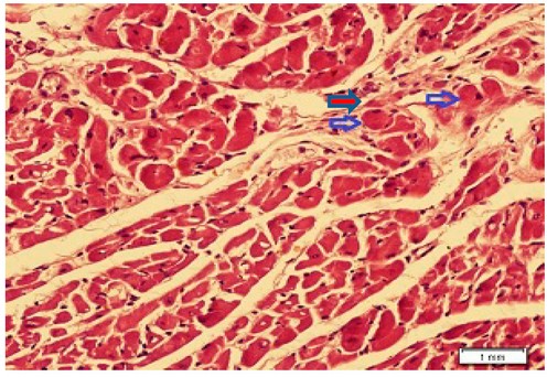
Figure 1. Shows heart tissue with degeneration of cardiomyocytes (blue arrow), disarrangement of cardiac muscle tissue and splitting of muscle bundles (red arrow) (H&E stain x400).
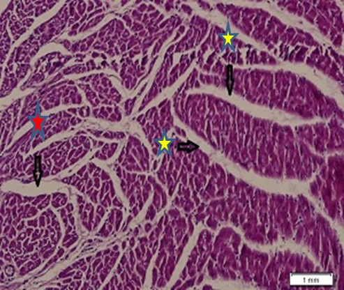
Figure 2. Shows heart tissue with marked degeneration of cardiomyocytes (yellow star) and dilated interstitial spaces (red star) (H&E stain x200).
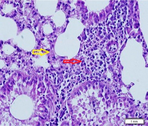
Figure 3. Showed lung tissue with marked lymphocyte infiltration (red arrow), edematous interstitial spaces and thickened alveolar walls (yellow arrow) (H&Ex400).
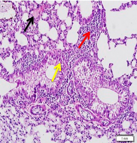
Figure 4. Shows lung tissue with edematous interstitial spaces and thickened alveolar walls (yellow arrow), marked lymphocytic infiltrate (red arrow) and congestion (black arrow) (H&Ex200).
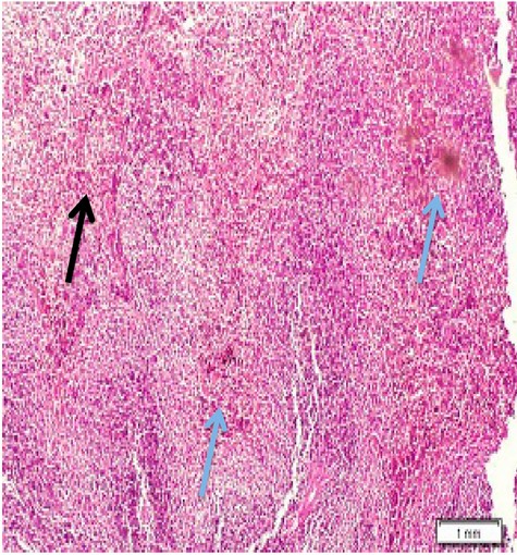
Figure 5. Shows spleen tissue with congestion( blue arrow) and a mild increase in red pulp area necrosis in white pulp (black arrow) (H&Ex100).
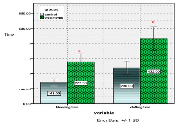
Figure 6. Showed bleeding, clotting time of treatment and control male mice after exposure to electromagnetic waves from mobile phone
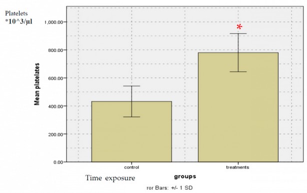
Figure 7. Showed platelets counts of treatment and control male mice after exposure to electromagnetic waves from mobile phone
*= significant differences (p≤0.0001).
DISCUSSION
Excessive exposure to (EMF) due to increased technologies becomes dangerous because of its effect on different organisms' biological systems and health. This research aimed to investigate histological disorders of some organs. The present study's result agrees with 19 who found damage in the heart tissue of mice exposed to the mobile phone (MP); this can be attributed to the use closely the heart can absorb EMR emitting from MP. In consonant with the present study, 20 found many disorders in the lung, such as infiltration of inflammatory cells, blood vessel obstruction and interalveolar septal disturbance. 21 Found exposure to EMR emitted from mobile can cause enlarged in the white pulps of the spleen and dilatation of its sinusoids; the degree of these changes increased with the duration of EMR exposure. Also, 22 found atrophy in seminiferous tubules and vacuolation in hepatocyte cells of guinea pigs exposed to EMW. The present study agreed with 23 when they discovered that many lesion in the tissue of brain rat exposed to EMW includes degeneration and edema, in lung found hemorrhage, emphysema and alveolar congestion. Atrophy with vacuolations in the hepatocyte, also necrosis in the pancreas. These changes can be attributed to many mechanisms: EMW with high energy causes increased local temperature leading to break down protein bound and denaturation 24. The second mechanisms where wave contact together to formed free radical production and antioxidant consumption which leads insufficient defense system, these free radicals attack lipids, proteins and nucleic acid, when causes genetic mutation leading to the breaking of DNA strands and cell death 25 26. Also a 900 MHz EMF application adversely influenced the learning behavior of female pups in the prenatal period and also resulted in histopathological changes occurring in the hippocampus 27. Histological changes can be due to the formation of free radicals through exposure to EMF, which in turn targets membrane lipids and changes their nature by breaking protein bonds 22. A study on workers, welders and computer operation exposed to EMW found increased RBC, MCV and platelets due to critical change in the erythropoiesis system 5. The present study, in agreement with 28, saw an increase in RBC, MCV and platelets. At the same time, a decrease in WBC, Hb and lymphocytes cell may be related to the effect of EMW on shortening cell cycles and increasing the synthesis of DNA. Another study found an increase in RBC and platelets correlated to the impact of EMW that causes stimulated division of stem cells in the bone marrow and increases immature reticulocyte cells 29. Results were found to increase bleeding and clotting time, and these deal with 12 who attributed these increase to the following reasons the first one, The increase in body temperature reduces blood viscosity, which eventually contributes to an increase in blood clotting time. The second, enzymatic chains or hormones experimentally affected the strength of the electromagnetic field can created around the outside of the cell wall and pull the ions to the opposite directions 30.
CONCLUSIONS
The effects of EMF on living tissues have been proven beyond a shadow of a doubt, and the level of damage is directly proportional to the magnetic field strength, time of exposure, and kind of tissue exposed.
Conflict of interest
no conflict of interest
Acknowledgment
We thank the Iraqi ministry of higher education and the University of Kerbala for the facilities needed to carry out this study. The authors personally funded this work.
Ethical approval:
The research related to human use has complied with all the relevant national regulations and institutional policies and, following the tenets of the Helsinki Declaration, has been approved by the author's institutional review board or equivalent committee. Project no. 1025 was approved on January 20th, 2019.
Conflict of interest:The authors declare they have no conflict of interest.
Informed consent statement: Informed consent has been obtained from all individuals included in this study.
REFERENCES
1. Odac\i E, \.Ikinci A, Y\ild\ir\im M, Kaya H, Akça M, Hanc\i H, et al. The effects of 900 megahertz electromagnetic field applied in the prenatal period on spinal cord morphology and motor behavior in female rat pups. NeuroQuantology. 2013;11(4):573–81.
2. Moussa EA. Effect of electromagenetic field on liver and kidney tissues of Swiss albino mice. JOURNAL-EGYPTIAN Ger Soc Zool. 2005;48(C):29.
3. Irmak MK, Fad\ill\io\uglu E, Güleç M, Erdo\ugan H, Ya\ugmurca M, Akyol Ö. Effects of electromagnetic radiation from a cellular telephone on the oxidant and antioxidant levels in rabbits. Cell Biochem Funct Cell Biochem its Modul by Act agents or Dis. 2002;20(4):279–83.
4. Leszczynski D, Joenväärä S, Reivinen J, Kuokka R. Non-thermal activation of the hsp27/p38MAPK stress pathway by mobile phone radiation in human endothelial cells: molecular mechanism for cancer-and blood-brain barrier-related effects. Differentiation. 2002;70(2–3):120–9.
5. Abdolmaleki A, Sanginabadi F, Rajabi A, Saberi R. The effect of electromagnetic waves exposure on blood parameters. Int J Hematol Stem Cell Res. 2012;13–6.
6. Funk RHW, Monsees T, Özkucur N. Electromagnetic effects--From cell biology to medicine. Prog Histochem Cytochem. 2009;43(4):177–264.
7. Velayutham P, Govindasamy GK, Raman R, Prepageran N, Ng KH. High-frequency hearing loss among mobile phone users. Indian J Otolaryngol Head Neck Surg. 2014;66(1):169–72.
8. Lippi G, Danese E, Brocco G, Gelati M, Salvagno GL, Montagnana M. Acute effects of 30 minutes of exposure to a smartphone call on in vitro platelet function. Blood Transfus. 2017;15(3):249.
9. Ng K-H. Non-ionizing radiations--sources, biological effects, emissions and exposures. In: Proceedings of the international conference on non-ionizing radiation at UNITEN. 2003. p. 1–16.
10. Heckenlively JR, Arden GB, Bach M. Principles and practice of clinical electrophysiology of vision. MIT press; 2006.
11. Iorio R, Delle Monache S, Bennato F, Di Bartolomeo C, Scrimaglio R, Cinque B, et al. Involvement of mitochondrial activity in mediating ELF-EMF stimulatory effect on human sperm motility. Bioelectromagnetics. 2011;32(1):15–27.
12. Ahmad DT. Effectes of Low Frequency Pulsed Magnetic Field on Blood Clotting Time in Male Rabbits. Diyala J Med. 2011;1(2):56–63.
13. Hua VM, Chen VMY. Procoagulant platelets and the pathways leading to cell death. In: Seminars in thrombosis and hemostasis. 2015. p. 405–12.
14. Van Poucke S, Stevens K, Marcus AE, Lancé M. Hypothermia: effects on platelet function and hemostasis. Thromb J. 2014;12(1):1–5.
15. Henriques U. Histological Technique in Routine Histopathology: An Opinion. Pathol Pract. 1981;171(3–4):417–22.
16. Rathkolb B, Fuchs H, Gailus-Durner V, Aigner B, Wolf E, de Angelis M. Blood collection from mice and hematological analyses on mouse blood. Curr Protoc Mouse Biol. 2013;3(2):101–19.
17. Brake MA, Ivanciu L, Maroney SA, Martinez ND, Mast AE, Westrick RJ. Assessing blood clotting and coagulation factors in mice. Curr Protoc Mouse Biol. 2019;9(2):e61.
18. Liu Y, Jennings NL, Dart AM, Du X-J. Standardizing a simpler, more sensitive and accurate tail bleeding assay in mice. World J Exp Med. 2012;2(2):30.
19. Ozguner F, Altinbas A, Ozaydin M, Dogan A, Vural H, Kisioglu AN, et al. Mobile phone-induced myocardial oxidative stress: protection by a novel antioxidant agent caffeic acid phenethyl ester. Toxicol Ind Health. 2005;21(7–8):223–30.
20. Hanafy LK, Karam SH, Saleh A. The adverse effects of mobile phone radiation on some visceral organs. Res J Med Med Sci. 2010;5(1):95–9.
21. Attia AA, Yehia MA. Histological, ultrastructural and immunohistochemical studies of the low frequency electromagnetic field effect on thymus, spleen and liver of albino swiss mice. Pak J Biol Sci. 2002;5(9):931–7.
22. Zare S, Alivandi S, Ebadi AG. Histological studies of the low frequency electromagnetic fields effect on liver, testes and kidney in guinea pig. World Appl Sci J. 2007;2(5):509–11.
23. Farahna M, Omer MAA, Garalnabi MEF, Al-Ganim AA, Abdelkareem S, Busharaa YM. The effects of static magnetic field on rats brain, lungs, liver, pancreas and blood electrolytes. NeuroQuantology. 2014;12(2).
24. McLauchlan KA. A possible mechanism for the effects of electromagnetic fields on biological cells. Elektor India Electron. 1993;11(3):47–9.
25. Balci M, Devrim E, Durak I. Effects of mobile phones on oxidant/antioxidant balance in cornea and lens of rats. Curr Eye Res. 2007;32(1):21–5.
26. Vecchia P, Matthes R, Ziegelberger G, Lin J, Saunders R, Swerdlow A. Exposure to high frequency electromagnetic fields, biological effects and health consequences (100 kHz-300 GHz). Int Comm Non-Ionizing Radiat Prot. 2009;378.
27. Ikinci A, Odaci E, Yildirim M, Kaya H, Akça M, Hanci H, et al. The effects of prenatal exposure to a 900 megahertz electromagnetic field on hippocampus morphology and learning behavior in rat pups. NeuroQuantology. 2013;11(4).
28. Antonopoulos A, Eisenbrandt H, Obe G. Effects of high-frequency electromagnetic fields on human lymphocytes in vitro. Mutat Res Toxicol Environ Mutagen. 1997;395(2–3):209–14.
29. Zeni O, Schiavoni A, Perrotta A, Forigo D, Deplano M, Scarfi MR. Evaluation of genotoxic effects in human leukocytes after in vitro exposure to 1950 MHz UMTS radiofrequency field. Bioelectromagn J Bioelectromagn Soc Soc Phys Regul Biol Med Eur Bioelectromagn Assoc. 2008;29(3):177–84.
30. Athanasiou A, Karkambounas S, Batistatou A, Lykoudis E, Katsaraki A, Kartsiouni T, et al. The effect of pulsed electromagnetic fields on secondary skin wound healing: an experimental study. Bioelectromagn J Bioelectromagn Soc Soc Phys Regul Biol Med Eur Bioelectromagn Assoc. 2007;28(5):362–8.
Received: January 25, 2022 / Accepted: October 22, 2022 / Published:15 November 2022
Citation: Yousif Al-Fatlawi A C. Evaluation of the effects of mobile phone electromagnetic radiation on some physiological parameters and histological structure in some laboratory male mice organs. Revis Bionatura 2022;7(4) 56. http://dx.doi.org/10.21931/RB/2022.07.04.56