2023.08.03.87
Files > Volume 8 > Vol 8 No 3 2023
Histomorphological study of tongue in Indigenous peacock (Pavo cristatus)

Hadaf H. Mohammed 1 and Luay O. Hamza 2*
1 Anatomy and Histology Department, Baghdad University, Iraq.
2 College of Veterinary Medicine, Baghdad University, Iraq.
*Corresponding author. [email protected]
Available from: http://dx.doi.org/10.21931/RB/2023.08.03.87
ABSTRACT
The current study aimed to examine the histomorphological features of the oral salivary ducts and the peacock tongue (Pavo cristatus) of this species. In this study, eight healthy adult peacocks were used after collecting them from a commercial market in Baghdad to investigate and describe the tongue in these bird species. After being euthanized, they were killed by cutting off the major neck blood vessels and bleeding until death; the tongue was identified. The tongue was the sharp end, elongated, somewhat triangular, and measured 8.2 mm. The apex, trunk, and base are its three sections. There are 4 to 5 layers of big conical papillae on the area where the root and body converge. Without gustatory papillae, the dense keratinized epithelium stratified squamous epithelia that lined the dorsal surface and were sparse on the ventral surfaces. The body and tongue root contain cells of the epithelium. It was supported by hyaline cartilage as a short plate. Lingual salivary glands situated anterior and posterior are superficial branch tubuloalveolar glands. This research was conducted at the University of Baghdad/ College of Veterinary Medicine in the Anatomy and Histology Department. Our result concluded that modifying the bird tongue, papillae distribution, and mucosa epithelium histological features is mainly restricted to feeding types that also have similarities with other avian species.
Keywords: peacock, tongue, lingual papillae, keratinized epithelium
INTRODUCTION
The indigenous peacock is the pheasant type native to Asian forests 1. There are three main species of the peacock, thought to have originated in Asia, Africa, and Australia. Many types of food in the bird nutrition, like plant seeds, flower heads and insects, also have been supplemented their diet by small mammals and reptiles to ensure that they are getting good nutrients 2. Peacocks are commonly found in dry savanna areas, deserts, and forests. Despite their large size, peacocks, in some situations, are capable of flight 1. Because of the different feeding habits, the tongues appear in various shapes and structures, especially in well-adapted foods 2, 3. The tongue of this bird differs from the tongues of mammals in that it has convex lateral portions, a middle sulcus, a variety of tongue papillae, and in some birds, an epithelium-coated apical dorsal surface 4, and the epithelium covers each dorsal and ventral surface of the tongue 5. This research aims to characterize the glands' spread and the mucosa's histomorphological structure in each section of the tongue.
MATERIALS AND METHODS
Eight healthy adult birds, both male and female, were obtained from a commercial market; they were euthanized by the intramuscular injection of a combination of diazepam and ketamine, then cutting off the major neck blood vessels and bleeding until death, the sample was washed by normal saline then collect eight tongues for the histological study, the samples were removed and immersed in 10% formalin. After fixation, the samples proceeded with the serial histological technique of blocks, then examined and analyzed by a light microscope; transversally and sagittal samples were cut and then prepared for paraffin embedding and 5-6 μm sections. Histologic samples were stained by hematoxylin-eosin 6, 7. The stained sections were examined by a light microscope and photographed with a digital camera.
RESULTS
The mature indigenous peacock tongue measures 8.2 millimeters in length, has a triangular form, and has an elongated terminating portion. The median fissure stretched along the body and root at the dorsal surface and was segmented into root, body, and tip. At the intersection of the body-base area across the root and body of the tongue, 4 to 5 massive conical papillae were oriented in that direction. The conical papillae were found on the lateral border (Fig.1).
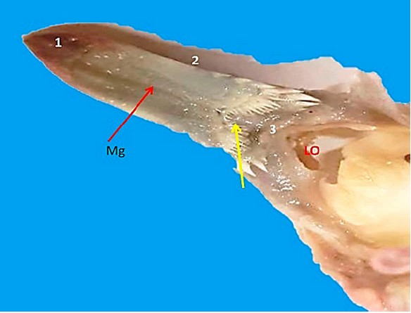
Figure 1. Dorsal view of the tongue: Apex (1), body(2). Lingual papillae (yellow arrow), median groove (Mg), and laryngeal opening (Lo)
Histologically, the peacock's tongue was enveloped by mucosa that varied depending on the location. Thick stratified squamous epithelium layers of stratum corneum covered the apex and dorsal surface of the tongue's body. In contrast, the ventral surface was either devoid of keratinized or thin epithelium. The covering of cornified cells has grown longer and compressed. Lamina propria and tunica submucosa are dense fibrous tissues containing lymphatic and blood arteries. Hyaline cartilage that reaches the tongue's tip supports the tongue's center (Fig. 2, 3, 4 & 5). The ventral, dorsal surface of the tongue body shows a modified structure represented by the scattered of many lymphatic nodules (Fig. 6, 7).
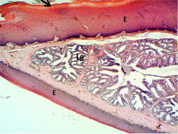
Figure 2. Histological section of the tongue showing keratinized stratified squamous epithelium (E), keratinized layer (black arrow), lingual salivary glands (lg).40x H&E stain
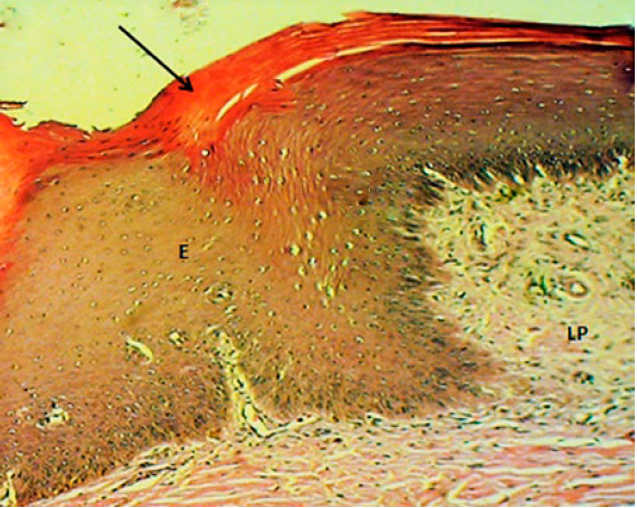
Figure 3. Microphotograph of the tongue tip (longitudinal section) showing thick stratified squamous epithelium (E), lamina propria (lp), and keratinized layer (black arrow).100x H&E stain
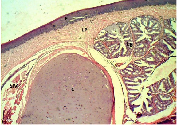
Figure 4. Microphotograph of the tongue (longitudinal section) showing epithelium(E), lamina propria (lp), lingual salivary glands (lg), hyaline cartilage(c) and skeletal muscle(SM). 40x H&E stain
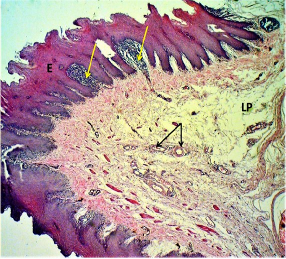
Figure 5. Microphotograph of the tongue dorsal surface showing epithelium(E), lamina propria (lp), blood vessels (black arrows), and lymphatic tissue (yellow arrows). 40x H&E stain
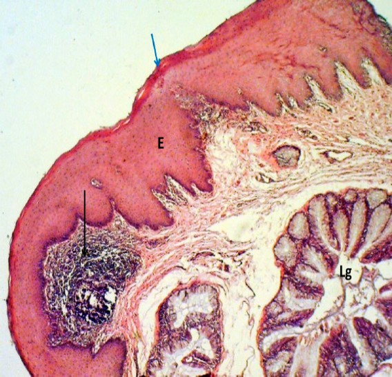
Figure 6. Microphotograph of the tongue dorsal surface showing epithelium(E), keratin layer (blue arrow), lymphatic nodule (black arrows) lingual salivary glands (lg). 100x H&E stain
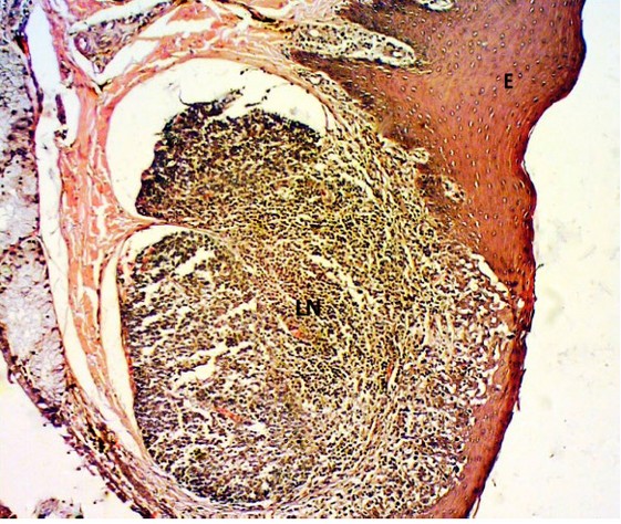
Figure 7 Microphotograph of the tongue (body) showing epithelium (E), lymphatic nodule (LN) 100x H&E stain.
In the mucosal lamina propria of the tongue, there are anterior and posterior glands located between the skeletal muscles; the results of glands revealed that these glands were tubule alveolar type, a connective tissue-thin layer sent trabeculae divided the gland into lobules, each lobule was contained mucous alveoli secreting, lined by cuboidal shape cells with nuclei located basally (Fig. 8, 9, 10, 11).
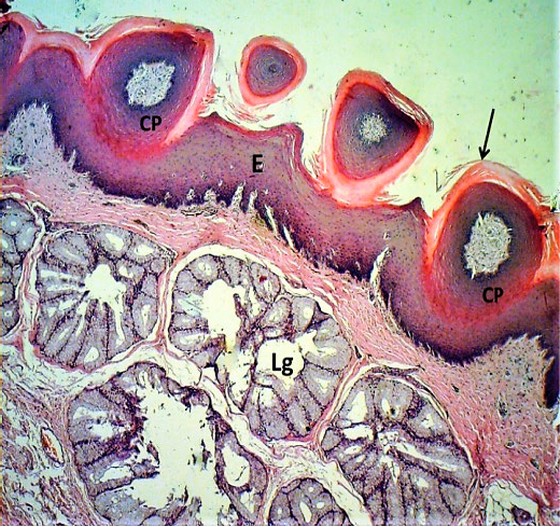
Figure 8. Microphotograph of the tongue dorsal surface showing epithelium (E), conical papillae (cp), keratinous layer (black arrows) lingual salivary glands (lg). 100x H&E stain
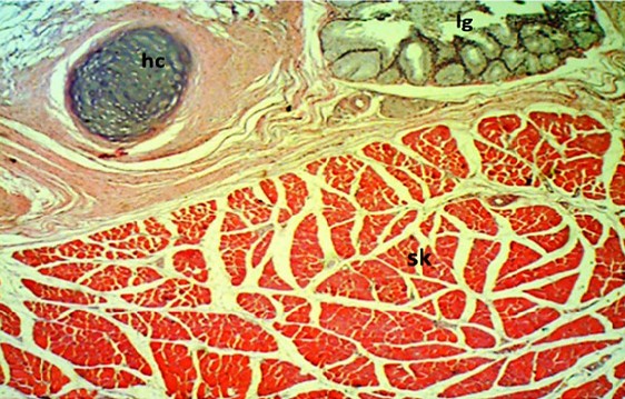
Figure 9. Microphotograph of the tongue (body) showing hyaline cartilage (HC), skeletal muscle (sk), anterior lingual glands (LG) 100x H&E stain
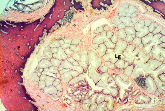
Figure 10. Microphotograph of the tongue (tongue base) showing: epithelium (E), simple branched tubule-alveolar mucous glands (lg). 100x H&E stain
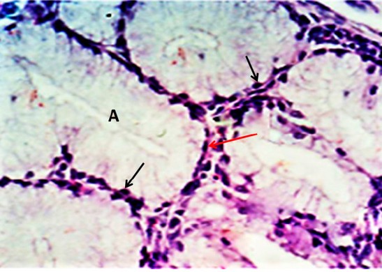
Figure 11. Magnified section of anterior lingual glands showing mucous alveoli lined by columnar cells with basal located nuclei (black arrows) and septa (red arrow). 400x H&E stain
DISCUSSION
The general structure of the tongue consists of three parts (root, body and free apex), which has 4 to 5 large conical papillae on the lateral border parallel with other observation by 8 that were recorded in avian quail and also been the same described in chicken 9. The way and the type of food intake made differences in the bird’s tongue structure while disagreeing with observation in carnivore birds like common kingfisher 10 Francolin 11.
The present result demonstrated that a tongue is shaped as triangular and contains many papillae located on different parts and surfaces of the tongue, the interior was covered by intrinsic lingual muscle and many conical papillae, similar observations have been recorded in some bird species like Egyptian goose and ostrich 12, 13.
It has been observed that the ventral surface of the tongue was devoid of keratinized epithelium or thin epithelium, while the dorsal surface and apex of the tongue were coated in dense keratinized stratified squamous epithelium; the same observation was found in common Myna and red jungle fowl14, 15. The thickness of keratinization in the epithelia depends on food intake; a high level of keratinization was observed in birds eating hard food, while low keratinization was shown in guinea fowl 16, 17.
On the other hand, the result showed that the dorsal and ventral surface of the tongue body has many scattered lymphatic nodules in dissimilar results of 18 in a chicken and 15 in a red jungle fowl while markedly noticed as that seen in other species like in Ostrich 19 that may be due to variation of species of birds.
There were numerous conical papillae on the dorsal surface of the tongue, which were of varying lengths and heights. The anterior and posterior surfaces of the conical papillae were coated with keratinized epithelia, As 20 had noted that a thin coating of keratinized stratified squamous epithelia coated the ventral and lateral surfaces of the tongue of Chukar partridge tongue. However, the type of food consumed significantly impacts the level of keratinization in tongue epithelia.
The lamina propria contains anterior and posterior glands, classified as a tubule alveolar type; it is divided into lobules that contain mucous alveoli secreting, this result was also mentioned in other species of birds like in the tongue of Chukar partridge also recognized in the study of the anterior salivary glands in chicken and partridge 20, 21. This result was in line with 13, who mentioned the location of the lingual gland in the dorsal and ventral surfaces of the ostrich tongue.
CONCLUSION
It was found in the current research that the distinctive histological features of the Peacock tongue, such as the presence of conical papillae encircled by a thick keratin layer and lymphatic nodules dispersed on the dorsal surface of the tongue, were present. Nevertheless, the findings could be used to clarify how diet affects this bird's lingual papillae's layout and form and how its salivary glands secrete.
REFERENCES
1. Gurjar RL, Singh RP, Mishra A. Density of the Indian Peafowl Pavo cristatus in Satpura Tiger Reserve, India. Journal homepage: www. wesca. net. 2013;8(1).
2. Mohammed HH. Prehatching Development of Gizzard in Indigenous Quail (Coturnixcoturnix japonica). Indian Journal of Natural Sciences. 2018; Vol.9 /Issue 51.
3. Mohammed HH. The Morphological and Histological Features of Tongue in Black Winged Kite (Elanus caeruleus). Basrah Journal of Veterinary Research. 2017;16(1):322-32.
4. Uppal V, Bansal N, Anuradha, Pathak D, Singh A. Light and scanning electron microscopy studies of quail tongues. Avian Biology Research. 2014 Aug;7(3):167-71.
5. Erdogan S, Sagsoz H, Akbalik ME. Anatomical and histological structure of the tongue and histochemical characteristics of the lingual salivary glands in the Chukar partridge (Alectoris chukar, Gray 1830). British Poultry Science. 2012 Jun 1;53(3):307-15.
6. Bancroft JD, Gamble M, editors. Theory and practice of histological techniques. Elsevier health sciences; 2008.
7. Hamza LO, Al-Mansor NA. Histological and histochemical observations of the small Intestine in the indigenous Gazelle (Gazella subgutturosa). J. Entomol. Zool. Studies. 2017;5(6):948-56.
8. Parchami A, Dehkordi RF, Bahadoran S. Fine structure of the dorsal lingual epithelium of the common quail (Coturnix coturnix). World Appl Sci J. 2010;10(10):1185-9.
9. Homberger DG, Meyers RA. Morphology of the lingual apparatus of the domestic chicken, Gallus gallus, with special attention to the structure of the fasciae. American Journal of Anatomy. 1989 Nov;186(3):217-57.
10. Al-Zahaby SA, Elsheikh EH. Ultramorphological and histological studies on the tongue of the common kingfisher in relation to its feeding habit. The Journal of Basic & Applied Zoology. 2014 May 1;67(3):91-9.
11. Kadhim KK, Atia MA, Hameed AT. Histomorphological and Histochemical Study on the Tongue of Black Francolin (" Francolinus francolinus"). International Journal of Animal and Veterinary Advances. 2014 Dec 20;6(6):156-61.
12. Hassan SM, Moussa EA, Cartwright AL. Variations by sex in anatomical and morphological features of the tongue of Egyptian goose (Alopochen aegyptiacus). Cells Tissues Organs. 2010;191(2):161-5.
13. Pasand AP, Tadjalli M, Mansouri H. Microscopic study on the tongue of male ostrich. Eur J Biol Sci. 2010;2(2):24-31.
14. Kadhim KK, Hameed AT, Abass TA. Histomorphological and histochemical observations of the Common Myna (Acridotheres tristis) tongue. International Scholarly Research Notices. 2013;2013.
15. Kadhim KK, Zuki AB, Babjee SM, Noordin MM, Zamri-Saad M. Morphological and histochemical observations of the red jungle fowl tongue Gallus gallus. African Journal of Biotechnology. 2011;10(48):9969-77.
16. Jackowiak H, Andrzejewski W, Godynicki S. Light and scanning electron microscopic study of the tongue in the cormorant Phalacrocorax carbo (Phalacrocoracidae, Aves). Zoological Science. 2006 Feb;23(2):161-7.
17. Venkatesan S, Shazia N, Kannan TA, Sabiha HB, Geetha R. Functional morphology of the epidermal structure of the feeding apparatus of guinea fowl (Numidameleagris). Int J Adv Res. 2015;3(10):1601-8.
18. Gargiulo AM, Lorvik S, Ceccarelli P, Pedini V. Histological and histochemical studies on the chicken lingual glands. British Poultry Science. 1991 Sep 1;32(4):693-702.
19. Jackowiak H, Ludwig M. Light and scanning electron microscopic study of the structure of the ostrich (Strutio camelus) tongue. Zoological Science. 2008 Feb;25(2):188-94.
20. Erdogan S, Sagsoz H, Akbalik ME. Anatomical and histological structure of the tongue and histochemical characteristics of the lingual salivary glands in the Chukar partridge (Alectoris chukar, Gray 1830). British Poultry Science. 2012 Jun 1;53(3):307-15.
21. Kudo KI, Nishimura S, Tabata S. Distribution of taste buds in layer‐type chickens: Scanning electron microscopic observations. Animal Science Journal. 2008 Dec;79(6):680-5.
Received: 25 June 2023/ Accepted: 26 August 2023 / Published:15 September 2023
Citation: Mohammed H H and Hamza L O. Histomorphological study of tongue in Indigenous peacock (Pavo cristatus). Revis Bionatura 2023;8 (3) 87 http://dx.doi.org/10.21931/RB/2023.08.03.87