2021.06.04.34
Files > Volume 6 > Vol 6 No 4 2021
Molecular Photoacoustic Imaging

Eduardo Cepeda1 and Katherine Narváez1
Available from http://dx.doi.org/10.21931/RB/2021.06.04.34
ABSTRACT
Medicine has gone through several challenges to make it much more accurate and thus prolong the human being's life. A large part of this challenge is diseased, so early detection can help carry out treatment on time. There is a technology that allows detecting an abnormality within the body without using an invasive method. Ultrasound is a diagnostic test used to scan organs and tissues through sound waves. Although this technique has been widely used, the results are not desired because the images generated are not high resolution.
On the other hand, X-rays are used because it presents an image with a much higher resolution than other techniques based on light waves or ultrasound; despite this, they are harmful to cells. In consequence of this problem, another method called molecular photoacoustic imaging has been implemented. This technique bridges the traditional depth limits of ballistic optical imaging and diffuse optical imaging's resolution limits, using the acoustic waves generated in response to laser light absorption, which has now shown potential for molecular imaging, allowing the visualization of biological processes in a non-invasive way. The purpose of this article is to give a critically scoped review of the physical, chemical, and biochemical characteristics of existing photoacoustic contrast agents, highlighting the pivotal applications and current challenges for molecular photoacoustic imaging.
Keywords. Photoacoustic Image, Biomedicine, Clinical Imaging, Optical Imaging.
INTRODUCTION
Optical imaging plays a crucial role in biomedicine because it provides a convenient way to visualize and understand biological events1, verifying the body's proper functioning and early detection of diseases2. Conventional images are known to have features that limit their ability to obtain in vivo images of tissues. Several widely used optical images are used, such as intrinsic signals3, magnetic resonance imaging (MRI)4, Spectroscopy5, laser staining contrast images6, and used. Optical images that use light to visualize cells have the characteristic that light undergoes a significant dispersion in biological tissue, which requires additional effort, so this method is relatively fast but limited7. Microscopy and other photon-using methods can provide high-resolution images. They achieve noise-free detection of individual absorbent nanoparticle shots, giving the technique a high potential for tissue cell applications, but only up to a depth of ~1mm in most of these biological tissues8.
MRI has been one of the highest resolution methods applied so far9. This technique manages to emit a 3D image10. It employs a mechanism in which the patient undergoes a magnetic field, so the protons, leading hydrogen, are aligned to this field, after which an electromagnetic pulse of radiofrequency is applied. This disturbs the balance of these atoms and introduces a transient phase of magnetization, which is perceived as a radio wave and transformed into an image11. One of its most significant limitations is that at the microscopic level is a deficiency in its detection sensitivity12.
Ultrasound images such as ultrasound, because they do not use radiation, are widely used in pregnant women13, which has had a high impact on producing two-dimensional up to three-dimensions ultrasound images14; this diagnostic test is based on emitting high-frequency sound waves. These waves travel through the body and bounce when colliding with density changes, allowing you to create an image15. Because they emit a longer wavelength and go de further into the tissues without dispersing in large magnitude, although the ultrasounds do not have a high resolution16. Computed tomography (CT) and fluorescence have always-on signals, and it is often challenging to design them to have biomarker-induced changes17. Also, they can provide depths of penetration by sacrificing spatial resolution18. The X-ray technique has been used to provide a clear image for diagnosing a fracture and detecting pneumonia19. This method consists of influencing a beam of X-rays into the body's tissues; this attenuation creates an overlapping shadow of the body region's internal structure to be studied. Thus a detector sensitive to X-rays transforms this transmitted fraction and converts it into an image16. Despite having a reasonably high resolution, the incidence of X-rays can cause mutations in fetuses, increasing the likelihood of developing cancer and cataracts, among other problems; that is why their exposure should be limited to the maximum18.
This method has different applications; it has been used to obtain preclinic images in vivo in small animals for various disease indicators. On the other hand, it has been applied significantly in cancer research: detection of primary tumors and molecular Characterization, therapeutic monitoring, identification, and evaluation of metastatic lymph nodes. This review focuses on molecular photoacoustic imaging (MPI), which has attracted increasing interest due to its specific advantages. This method allows to visualize and quantify biological processes at the molecular and cellular level in a non-invasive way, providing an opportunity to detect, stage, predict, and monitor diseases' development.
Methods for MPI development
MPI is a pop-up method that combines the optical image's high contrast with the ultrasound's high spatial resolution. The success of the MPI is based on the intrinsic properties of this physical process20. During this procedure, the photo energy influenced by short laser pulses is absorbed by contrast agents, either exogenous or endogenous, partially converted into heat, resulting in increased broadband acoustic waves at MHz frequencies21. These waves can be detected by an ultrasound transducer on the tissue surface and reconstructed to form an image of the absorbed optical energy distribution and, therefore, the formation of acoustic photo imaging22.
Obtaining MPI relies on exogenous or endogenous contrast agents to transform absorbed photons into heat. It is not invasive for the in vivo organism. Various inorganic nanomaterials have been shown, such as quantum dots (QD), carbon nanotubes, gold nanoparticles, and silver nanoplates23. They are promising as contrast agents in MPI. Also, as sound waves are less dispersed in tissue than photons, MPI can overcome the limitations of traditional optical images. Some MPI methods have been developed to obtain images of biological tissues such as MPI using endogenous chromophores like hemoglobin, melanin, water or lipids 24, MPI using dyes25, MPI with nanostructures like silver nanoplates 23, and molecular photoacoustic contrast agents (MPCA)26. The use of the different contrast agents will depend on the depth you want to reach at the study time. MPI can reach a penetration depth of several cm with an order resolution of about 100um27. The perspective provides an overview of each method mentioned to help and provide valuable and current MPI information for future applications such as cancer.
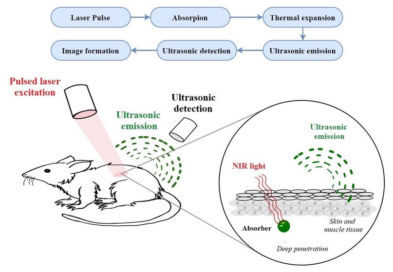
Figure 1. Schematic illustration, which shows the process of MPI.
Contrast agents
As mentioned above, a throbbing laser is used, and this range's depth will depend on its wavelength. Contrast agents absorb the laser to generate MPI, which can be endogenous or exogenous process28. These agents must possess three physical photo properties: Their maximum absorbance wavelength must be between 680-950nm. To obtain a higher photoacoustic signal, Quantum fluorescence performance should be low to maximize energy dissipated through non-radioactive pathways. Another important property is that its extinction coefficient is higher than 104 M-1cm-1 to maximize the amount of light absorbed29.
Endogenous
There are endogenous contrast agents, i.e., produced by the body, including water and lipids, which are weak chromophores compared to hemoglobin, a protein of 64kDa, absorbs much more than the chromophores present in other tissues, has a wavelength range between 950nm-1400nm oxygenated and deoxygenated28. Water has absorption bands of 970nm, 1200nm, and <1400nm, while lipids are at wavelengths of 930nm, 1040nm, 1210nm, and 1390nm. Although endogenous chromophores have long wavelengths, they need exogenous contrast agents for MPI to have a high resolution30.
Exogenous
Exogenous contrast agents must comply with specific properties. Physical photo properties: high molar coefficient of extinction to maximize the amount of light absorbed; characteristic absorption spectrum to avoid confusion, even at low concentrations; have a wavelength between 650nm-950nm, among others31. Biological properties: orientation and biocompatibility must overcome cellular barriers, the size of the targeting molecules must be small to cross physiological barriers32. There are different types of exogenous chromophores, such as:
Nanostructures
Nanostructures have also been a method used as contrast agents in MPI. Combining optical images such as MPI is very useful for therapeutic tracking, thus having the potential to propel the nanomedicine field towards authentic personalized medicine33. There are two main classifications according to their physical properties. The first group is based on surface plasmon resonance (SPR), specific property of certain metals such as gold3435. This property occurs when the surface-free loads of these nanoparticles oscillate with an electromagnetic field, leading to optical absorption 36. The second group is dyed-containing nanoparticles.
An example is a water-soluble indocyanine. Most of these contrast agents are encapsulated in layers of nanoparticles 36. The nanoparticles used have an absorption peak in the NIR region because tissue attenuation is lower at 37.
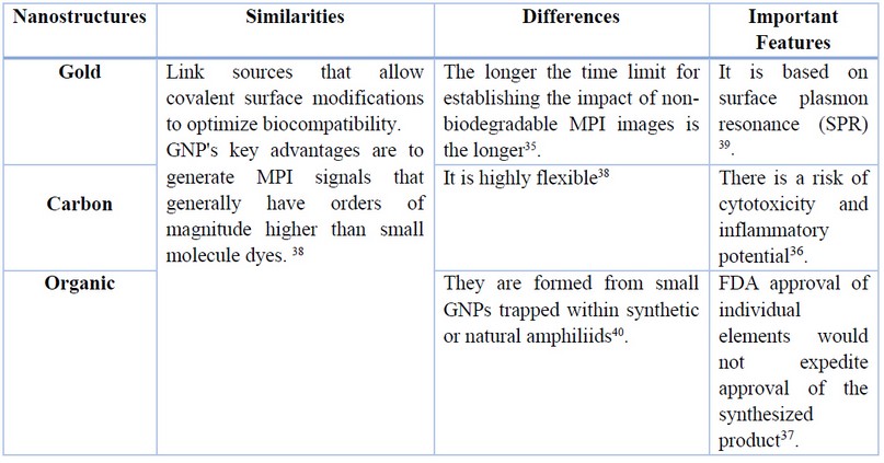
Table 1. Essential features of the most commonly used nanostructures for MPI applications.
Molecular Photoacoustic Contrast Agents
MPCA can be classified into three types: Linear absorption (LA)41: In this case, the dye has a shorter excitation state than the laser pulse. There is no absorption in that state, and a linear dependence on the amplitude of the PA signal is observed. Saturable absorber (SA)42: Its absorption in a state of excitation is negligible, but its lifespan is much longer than the laser pulse. Reverse-saturable absorber (RSA)41: In this type of canning agent, a nonlinear increase in absorption and PA response is observed by increasing laser creep43.
Cyanine dyes
Cyanine dyes are an exogenous contrast agent. Two halves of nitrogen, indolin heterocycles, thiazole, or quinoline joined by a linear polymethine chain, may be composed of 1, 3, 5, or 7 carbons44. Water-soluble indocyanine (ICG) has been extensively studied for in vivo fluorescence imaging due to its low toxicity 43. FDA approved the use of this contrast agent with nanoparticle encapsulation. Cyanine dyes have a molar extinction coefficient in a range between a Furthermore, quantum fluorescence performance (proportion of photons emitted relative to the number of photons absorbed) in a range of 11.30% to 4.39%45.
Curcumin dyes
This dye is found in nature in the rhizomes of Curcuma plants46. This plant is known to possess anti-inflammatory, purifying, antifungal, antibacterial properties 47. The boron difluoride derivative of natural curcumin (curcumin BF2) is a compound with a high quantum fluorescence performance, which may exhibit an amplified photoacoustic contrast to the standard crystal violet compound has been widely used in molecular photoacoustic images48. Curcumin BF2 has a maximum wavelength of 498nm. Because this wavelength is less than recommended, a 4-dimethylaminophenyl group is introduced at the end ends of the main chain to increase its wavelength to 684nm49.
BODIPY
BODIPY contrast agents have been widely used as signaling molecules and in imaging50. Despite this, nude BODIPY chromophores cannot be used in MPI because they have very high fluorescence. In this way, they have been combined with 1H-pyrrole (PyBODIPY) and PEG-400 to improve aqueous solubility to apply this compound in vivo imaging. These contrast agents' most important properties are: wavelength greater than 800nm, are non-photo-toxic, photo-stable, and have a high extinction coefficient51.
Biomedical applications
Molecular photoacoustic imaging is a technique that has various biomedical applications thanks to its advantages over other imaging methods. This technique is safe and effective in diagnosing diseases by providing images of different tissues' morphological structure and physiological characteristics. It has also been proposed as a tool to guide in vivo therapies5253.
Cancer Imaging
MPI is a non-ionizing technique that can be captured in real-time54. For cancer screening, it is necessary to locate which regions of the body are infected with tumors. Because a tumor is a buildup of tissue, which has cells that undergo abnormal growth, this formation also needs nutrients that will be transported by blood; As mentioned above, hemoglobin is a dominant endogenous contrast agent in the optical window so, high contrast images of the microvasculature can be obtained around the tumor54. Although hemoglobin is a potent contrast agent, it is necessary to use various exogenous chromophores; because tumors have leaky vascular systems, a low lymphatic drainage system, nanometer-sized contrast agents conjugated with targeted ligands such as peptides, antibodies have been used to bind to receptors that are in an over-exposed form in tumor tissue; with it, you get an image55.
Imaging of Atherosclerosis Plaques
Atheroma plaques are an injury that affects the cardiovascular system, and these appear in the body due to the accumulation of low-density cholesterol (LDL), which causes the radius of the arteries. In some cases, the artery is so affected that it can explode from a lack of diagnosis. This has been one of the biggest causes of death in industrialized countries54, in which the population tends to eat junk food. There are other methods for detecting atheroma plaques, such as angiography or ultrasound. However, they do not provide sufficient information. Also, angiography uses x-rays, so this study should be minimized to the maximum because it can be harmful to the patient. Intravascular and extravascular molecular photoacoustic imaging (IVPA) and extravascular (EVPA) have been shown to detect atheroma plaques thanks to their composition54. Endogenous chromophores have been shown to contrast specific components present in this atheroma plaque, such as lipids, calcium deposits, macrophage content, and fibrous material; However, it is necessary to use exogenous contrast agents directed with biomarkers to intensify differentiation and improve image quality549.
Similarly, MPI has been used to obtain stent images of the coronary arteries. This technique applies to arteries that have a severe case of atherosclerosis. Although stents are successful, they can bring restenosis and hyperplasia, so to treat it, you need imaging. In these cases, IVPA is used because it shows good penetration into the tissue and a high resolution. These images are taken ex vivo. Other methods are used to obtain in vivo images, but the stent is made of metal, which is very susceptible and challenging to obtain the image53.
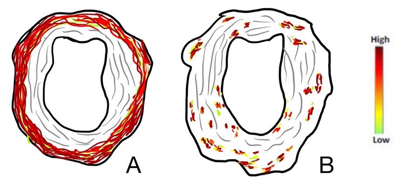
Figure 2. Ultrasound and photoacoustic imaging of an atherosclerotic rabbit aorta.
CONCLUSION
MPI is a modality with increasing interest that allows simultaneous imaging through endogenous chromophores present in exogenous tissues and contrast agents that visualize biological processes in vivo. These chromophores are of paramount importance in applying molecular photoacoustic images because they are responsible for absorbing energy. Compared to traditional optical images, molecular acoustic photo images have advantages because they detect acoustic signals that are much less attenuated than photons in tissues. In addition to using non-ionizing radiation and real-time images with high spatial resolution, technology also can perform a relatively inexpensive system. Due to these advantages, this molecular imaging modality has allowed the detection of biological and pathological events in vivo at unprecedented tissue depths with enhanced fluorescence images that traditional optical images expose us to. For this reason, molecular acoustic photo images are in high demand due to their relatively high advantage and biosecurity in living organisms.
This method is expected to significantly improve deep tissue images of anatomical structures, disease-related biomarkers, or physiological processes to improve the diagnosis of life-threatening diseases. In addition to obtaining deep tissue images, the flexibility of this method opens up doors of integration with other functional remains for multiple purposes, such as multifunctional theragnostic platforms, studies in oncology, neuroscience, and cardiovascular diseases; preliminary clinical trials have focused mainly today on the detection, staging and therapeutic follow-up of cancer.
Competing interest
The authors declare that they have no competing interests
REFERENCES
1. Wang, S., Larina, I. V. & Larin, K. V. Label-free optical imaging in developmental biology [Invited]. Biomed. Opt. Express 11, 2017 (2020).
2. Godavarty, A., Rodriguez, S., Jung, Y. J. & Gonzalez, S. Optical imaging for breast cancer prescreening. Breast Cancer Targets Ther. 7, 193–209 (2015).
3. Begum, M., Joiner, D. P. & Ts'o, D. Y. Stimulus-driven retinal intrinsic signal optical imaging in mouse demonstrates a dominant rod-driven component. Investig. Ophthalmol. Vis. Sci. 61, (2020).
4. Wu, L. et al. Perfluorocarbons-Based Imaging in Biomedicine. 7377–7395 (2020).
5. Steinegger, A., Wolfbeis, O. S. & Borisov, S. M. Optical Sensing and Imaging of pH Values: Spectroscopies, Materials, and Applications. Chem. Rev. (2020) doi:10.1021/acs.chemrev.0c00451.
6. Iancu, S. D. et al. Assessment of gold-coated iron oxide nanoparticles as negative T2 contrast agent in small animal MRI studies. Int. J. Nanomedicine 15, 4811–4824 (2020).
7. Kopek, B. G. et al. Diverse protocols for correlative super-resolution fluorescence imaging and electron microscopy of chemically fixed samples. Nat. Protoc. 12, 916–946 (2017).
8. Adhikari, S. et al. Photothermal Microscopy: Imaging the Optical Absorption of Single Nanoparticles and Single Molecules. ACS Nano (2020) doi:10.1021/acsnano.0c07638.
9. Rich, L. J. et al. 1H magnetic resonance spectroscopy of 2H-to-1H exchange quantifies the dynamics of cellular metabolism in vivo. Nat. Biomed. Eng. 4, 335–342 (2020).
10. Watson, W. D. et al. Use of cardiac magnetic resonance to detect changes in metabolism in heart failure. Cardiovasc. Diagn. Ther. 10, 583–597 (2020).
11. Cohen, M. S. & Bookheimer, S. Y. Localization of brain function using magnetic resonance imaging. Trends Neurosci. 17, 268–277 (1994).
12. Degen, C. L., Poggio, M., Mamin, H. J., Rettner, C. T. & Rugar, D. Nanoscale magnetic resonance imaging. Proc. Natl. Acad. Sci. U. S. A. 106, 1313–1317 (2009).
13. Edzie, E. K. M. et al. Perception of Ghanaian Primigravidas Undergoing Their First Antenatal Ultrasonography in Cape Coast. Radiol. Res. Pract. 2020, 1–10 (2020).
14. Hu, J. Q. et al. Application of two-dimensional and three-dimensional ultrasound in prenatal screening for brachydactyly deformity. Am. J. Transl. Res. 12, 5827–5835 (2020).
15. Hu, J. et al. Diagnosis of liver tumors by multimodal ultrasound imaging. Medicine (Baltimore). 99, e21652 (2020).
16. Seibert, J. A. Part 1: Basic principles of x-ray production. J Nucl Med Technol 32, 139–47 (2004).
17. Kounatidis, I. et al. 3D Correlative Cryo-Structured Illumination Fluorescence and Soft X-ray Microscopy Elucidates Reovirus Intracellular Release Pathway. Cell 182, 515-530.e17 (2020).
18. Palermo, F. et al. X-ray Phase Contrast Tomography Serves Preclinical Investigation of Neurodegenerative Diseases. Front. Neurosci. 14, 1–11 (2020).
19. Since January 2020 Elsevier has created a COVID-19 resource centre with free information in English and Mandarin on the novel coronavirus COVID- 19 . The COVID-19 resource centre is hosted on Elsevier Connect , the company' s public news and information . (2020).
20. Frigenti, G. et al. Microbubble resonators for all-optical photoacoustics of flowing contrast agents. Sensors (Switzerland) 20, 1–10 (2020).
21. Mooney, T. A. et al. Listening forward: Approaching marine biodiversity assessments using acoustic methods: Acoustic diversity and biodiversity. R. Soc. Open Sci. 7, (2020).
22. Lediju Bell, M. A. Photoacoustic imaging for surgical guidance: Principles, applications, and outlook. J. Appl. Phys. 128, 1–13 (2020).
23. Park, B. et al. Deep tissue photoacoustic imaging of nickel(II) dithiolene-containing polymeric nanoparticles in the second near-infrared window. Theranostics 10, 2509–2521 (2020).
24. Graham, M. T., Huang, J., Creighton, F. X. & Lediju Bell, M. A. Simulations and human cadaver head studies to identify optimal acoustic receiver locations for minimally invasive photoacoustic-guided neurosurgery. Photoacoustics 19, 100183 (2020).
25. Ollé, E. P., Farré-Lladós, J. & Casals-Terré, J. Advancements in microfabricated gas sensors and microanalytical tools for the sensitive and selective detection of odors. Sensors (Switzerland) 20, 1–39 (2020).
26. Tsang, V. T. C., Li, X. & Wong, T. T. W. A review of endogenous and exogenous contrast agents used in photoacoustic tomography with different sensing configurations. Sensors (Switzerland) 20, 1–20 (2020).
27. Gonzalez, E. A. & Bell, M. A. L. GPU implementation of photoacoustic short-lag spatial coherence imaging for improved image-guided interventions. J. Biomed. Opt. 25, 1 (2020).
28. Beard, P. Biomedical photoacoustic imaging. Interface Focus 1, 602–631 (2011).
29. Knox, H. J. & Chan, J. Acoustogenic Probes: A New Frontier in Photoacoustic Imaging. Acc. Chem. Res. 51, 2897–2905 (2018).
30. Cao, Q., Zhegalova, N. G., Wang, S. T., Akers, W. J. & Berezin, M. Y. Multispectral imaging in the extended near-infrared window based on endogenous chromophores. J. Biomed. Opt. 18, 101318 (2013).
31. Li, T. et al. The novel DPP-BDT nanoparticles as efficient photoacoustic imaging and positron emission tomography agents in living mice. Int. J. Nanomedicine 15, 5017–5026 (2020).
32. Future Potential. Outsourcing to India 281–291 (2005) doi:10.1007/3-540-24794-7_25.
33. Tabish, T. A. et al. Smart Gold Nanostructures for Light Mediated Cancer Theranostics: Combining Optical Diagnostics with Photothermal Therapy. Adv. Sci. 7, 1–28 (2020).
34. Yang, X., Stein, E. W., Ashkenazi, S. & Wang, L. V. Nanoparticles for photoacoustic imaging. Wiley Interdiscip. Rev. Nanomedicine Nanobiotechnology 1, 360–368 (2009).
35. Gandolfi, M., Banfi, F. & Glorieux, C. Optical wavelength dependence of photoacoustic signal of gold nanofluid. Photoacoustics 20, (2020).
36. Li, W. & Chen, X. Gold nanoparticles for photoacoustic imaging. Nanomedicine 10, 299–320 (2015).
37. Ge, X. et al. Light-activated gold nanorod vesicles with NIR-II fluorescence and photoacoustic imaging performances for cancer theranostics. Theranostics 10, 4809–4821 (2020).
38. Gerosa, C. et al. Gold nanoparticles: A new golden Era in oncology? Pharmaceuticals 13, 1–18 (2020).
39. Gil-Moles, M. et al. Gold Metallodrugs to Target Coronavirus Proteins: Inhibitory Effects on the Spike-ACE2 Interaction and on PLpro Protease Activity by Auranofin and Gold Organometallics**. Chem. - A Eur. J. 26, 15140–15144 (2020).
40. Sarraf, M., Nasiri-tabrizi, B. & Hong, C. Since January 2020 Elsevier has created a COVID-19 resource centre with free information in English and Mandarin on the novel coronavirus COVID- 19 . The COVID-19 resource centre is hosted on Elsevier Connect , the company' s public news and information . (2020).
41. Liu, L. et al. Highly sensitive broadband differential infrared photoacoustic spectroscopy with wavelet denoising algorithm for trace gas detection. Photoacoustics 21, 100228 (2021).
42. Turani-i-Belloto, A., Brunet, T., Khaldi, A. & Leng, J. A sacrificial route for soft porous polymers synthesized via frontal photo-polymerization. Polymers (Basel). 12, (2020).
43. Borg, R. E. & Rochford, J. Molecular Photoacoustic Contrast Agents: Design Principles & Applications. Photochem. Photobiol. 94, 1175–1209 (2018).
44. Wang, Y. et al. Targeted nanobubbles carrying indocyanine green for ultrasound, photoacoustic and fluorescence imaging of prostate cancer. Int. J. Nanomedicine 15, 4289–4309 (2020).
45. Mu, H. et al. pH-Activatable Cyanine Dyes for Selective Tumor Imaging Using Near-Infrared Fluorescence and Photoacoustic Modalities. ACS Sensors (2020) doi:10.1021/acssensors.0c01926.
46. Sharifi-Rad, J. et al. Turmeric and Its Major Compound Curcumin on Health: Bioactive Effects and Safety Profiles for Food, Pharmaceutical, Biotechnological and Medicinal Applications. Front. Pharmacol. 11, 1–23 (2020).
47. Khattak, S., Saeed-ur-Rehman, Shah, H. U., Ahmad, W. & Ahmad, M. Biological effects of indigenous medicinal plants Curcuma longa and Alpinia galanga. Fitoterapia 76, 254–257 (2005).
48. Bellinger, S. et al. Characterization of a NIR absorbing thienyl curcumin contrast agent for photoacoustic imaging. Chem. Commun. 54, 6352–6355 (2018).
49. Frenette, M. et al. Shining light on the dark side of imaging: Excited state absorption enhancement of a bis-styryl bodipy photoacoustic contrast agent. J. Am. Chem. Soc. 136, 15853–15856 (2014).
50. Fan, Y. et al. Architectures and Applications of BODIPY-Based Conjugated Polymers. Polymers (Basel). 13, 75 (2020).
51. Merkes, J. M. et al. Tuning Optical Properties of BODIPY Dyes by Pyrrole Conjugation for Photoacoustic Imaging. Adv. Opt. Mater. 8, 1–9 (2020).
52. Fu, Q., Zhu, R., Song, J., Yang, H. & Chen, X. Photoacoustic Imaging: Contrast Agents and Their Biomedical Applications. Adv. Mater. 31, (2019).
53. Hoda Badr, Cindy L. Carmack, Deborah A. Kashy, Massimo Cristofanilli, and T. A. R. 基因的改变NIH Public Access. Bone 23, 1–7 (2011).
54. Luke, G. P., Yeager, D. & Emelianov, S. Y. Biomedical applications of photoacoustic imaging with exogenous contrast agents. Ann. Biomed. Eng. 40, 422–437 (2012).
55. Wang, S., Lin, J., Wang, T., Chen, X. & Huang, P. Recent advances in photoacoustic imaging for deep-tissue biomedical applications. Theranostics 6, 2394–2413 (2016).
Received: 15 June 2021
Accepted: 10 September 2021
Eduardo Cepeda1 and Katherine Narváez1
1. School of Biological Sciences and Engineering, Yachay Tech University, Urcuquí 100650, Ecuador;
Corresponding author: [email protected]; [email protected]
ORCID
Eduardo Cepeda https://orcid.org/0000-0002-4074-6558
Katherine Narváez https://orcid.org/0000-0003-3772-2018