2022.07.03.37
Files > Volume 7 > Vol 7 No 3 2022
A Comparative Histomorphological and Histochemical Study of the Ventriculus between Iraqi Adult Geese (Anser anser) and Guinea fowls (Numidia meleagris)

Iman Mousa Khaleel 1* ,Khalid Ibrahim Abd Alkhazraji2 ,
1,Department of Anatomy and Histology, College of Veterinary Medicine, University of Baghdad, Iraq
2Department of Anatomy and Histology, College of Veterinary Medicine, University of Diyala, Iraq
Corresponding author: [email protected],
Available from: http://dx.doi.org/10.21931/RB/2022.07.03.37
ABSTRACT
The present study aimed to describe and compare the histomorphological and histochemical structures to ventriculus in Goose(Anser anser) and Guinea fowls (Numidia meleagris). The work was carried out on twenty apparently clinically healthy birds obtained from a supplier in Baghdad city. They were allocated in two equal groups of each type of bird. The two groups were utilized for histological and histochemical investigations of their ventriculus organ. After anesthesia and killing birds, their abdominal cavity was dissected, their ventriculus was identified and proper specimens from its walls were prepared. The samples designated for histochemical staining were fixed in a solution of bruin's fixative, while the others for general histological examination were fixed in (10%) neutral buffered formalin. After processing, the sections were stained with (2.5 PH) Alcian- PAS combination, periodic acid Schiff, Masson Trichrome, and Hematoxylin and Eosin stains. This study elucidates that the microscopic construct of the ventriculus was similar for the two species.
The ventriculus consists of three tunicates (serosa, muscularis, and mucosa), whereas the submucosa is absent. The wall showed some significant differences in morphometric measurements. The mucosa of the ventriculus is coated with a thick layer of cuticles organized as a wavy clear pink line parallel to the mucosal surface and mucosal folds. The simple cuboidal-columnar epithelium covered the mucosa, which showed many folds. The proprial glands (simple tubular type) are lined by simple cuboidal epithelium, which opens into the crypts between the folds. A well-developed muscular comprised of smooth muscle fibers as two layers of inner circular and outer longitudinal. Serosa is composed of loose connective tissue coated by mesothelium. The Mean thickness of Cuticle, length of folds, mucosa and muscular in goose were significantly higher than that in guinea fowl; these differences may be due to variation in their diet. PAS and AB-PAS combined (2.5-PH) stains, cuticle layer, epithelium lining, and gastric glands showed a positive reaction with these stains. This study aimed to Study the normal histomorphological histochemical and structure of Gizzard in two avian species, local male guinea fowl (Numidia meleagris) and male geese (Anser anser). Also aimed to Comparative histomorphological, histomorphometric measurements and histochemical study of the Gizzard of two different local male avian species (Guinea fowl and geese).
Keywords: Histomorphological, Histochemical, Ventriculus, Guinea fowls, Geese
INTRODUCTION
Avian species have different adaptations for the digestive processing of their foods1. Several kinds of research were designed on habits of food and affecting activity and characteristics of morphology in avian digestion organs2. The digestive organs treat as machines that act for maintaining numerous activities; therefore, the differences in their functional capacities may affect their metabolic rates. Aves have high metabolic rates, which correspond with their requirement; thus, they must eat a large amount of diet 3. Generally, the stomach is considered the most active portion of the avian digestive system; it consists of two parts, that are the proventriculus and ventriculus or Gizzard. The shape and size of these two parts of the avian stomach were varied and affected by the type of food. The proventriculus wall was- thin and glandular 4. The ventriculus contains grit, which facilitates beside muscle in grinding up the foods5. Birds were divided into three types according to their stomach type eating birds in which the ventriculus has mechanical action on a diet, such as a sparrow and a turkey, secondly the soft eating birds in which the ventriculus (Gizzard) as a stockist of food represented by owl and kestrel intermediate food eating birds as in hoopoe and geese in which the main function of ventriculus(Gizzard) was storage and physical digestion6.
MATERIALS AND METHODS
Birds
The current study was conducted on ten healthy male birds, guinea fowl and ten male goose obtained from a local Baghdad city supplier. This study was done in the laboratory of the Department of Anatomy and Histology, College of Veterinary Medicine, University of Baghdad.
Histological Technique
Specimens from ventriculus tissue were taken from a different region, washed with normal saline and fixed in 10% buffered formalin saline. All samples were processed in a series concentration of ethanol alcohol procedure and section at 6-7µm. Some sections were stained routinely with hematoxylin and eosin to detect the construction of the studied tissue; others with special stain Masson's Trichrom for collagen and smooth muscle fibers. Periodic acid Schiff (PAS) Stain was used for mucopolysaccharides, while PAS-AB(PH 2.5) for neutral and acidic mucins7. The histological sections were photographed using a digital camera connected to a computer; sections were photographed at different adjustment power(3.7 x,4x,10x and 40x ).
Statistical Analysis
The Statistical Analysis System- SAS 8 program, Users Guide. (Statistical Version 9.1th ed SAS. (USA).
RESULTS
The microscopic observations of the ventriculus in male goose and guinea fowl generally were similar, which revealed that the ventricular wall in both species was comprised of tunica serosa, muscularis, submucosa not present and mucosa. The mucous membrane in the ventriculus was covered by a layer of thick Keratin-like secretion known (Cuticle) which was organized as a wavy clear pink line parallel to the mucosal surface and the mucosal folds (Fig.1) . Mean thickness of this layer was significantly higher in geese than in guinea fowls (Table1).
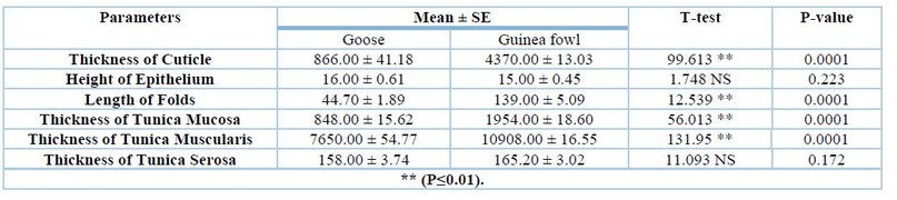
Table 1. Comparison between Adult Male Guinea fowls and Geese in parameters study
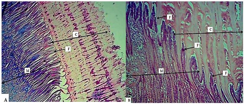
Figure 1. Photomicrograph illustrate the Ventricular wall showed: Cuticle (C), Mucosa ( M)Mucosal folds(F). A- 10 X Masson's TrichTrichrome Stain in Guinea fowl. B- X 10 H&E Stain in Goose.
The mucosa was thrown into distinct numerous narrow longitudinal mucosal folds, which lining mostly with simple cuboidal to columnar cells (Fig.2). Proprial glands (tubular mucous glands) were lined by simple cuboidal epithelium(Fig.3). The mean length of folds and mean thickness of tunica mucosa was significantly higher in geese than in guinea fowls, but non especially difference in the height of lining epithelium between the two species (Table1).
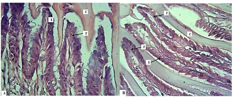
Figure 2. A: Photomicrograph illustrates the Ventricular showed: Epithelium (E), fold (F), Cuticle(C), A- X 40 H&E in Goose, B- X 40 H&E Stain in Guinea f
The Propecia was filled by gastric or proprial glands (simple tubular glands), which opened into the crypts between the folds and arranged into two portions, the first were long, straight, and its contents were ejected, while the second one was short, oval-shaped and packed, the proprial connective tissue observed extended into the core of the narrow fold (Fig.3,4), also the propria was wealthy by the collagenous fibers. The muscularis mucosa was present but interrupted in the ventriculus wall; the submucosal layer was absent (Fig. 5).
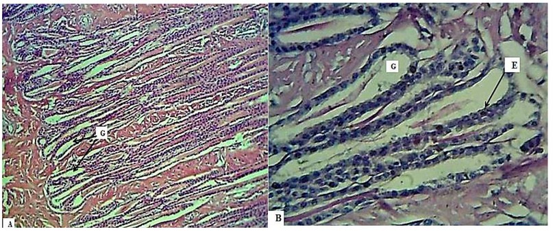
Figure 3. Photomicrograph: illustrate the Ventricular showed: Mucosal or proprial Glands(G), Epithelium; Simple cuboidal epithelium (E), A-10X H&E Stain in Male Goose. B- 40 X PAS Stain in Male Goose.
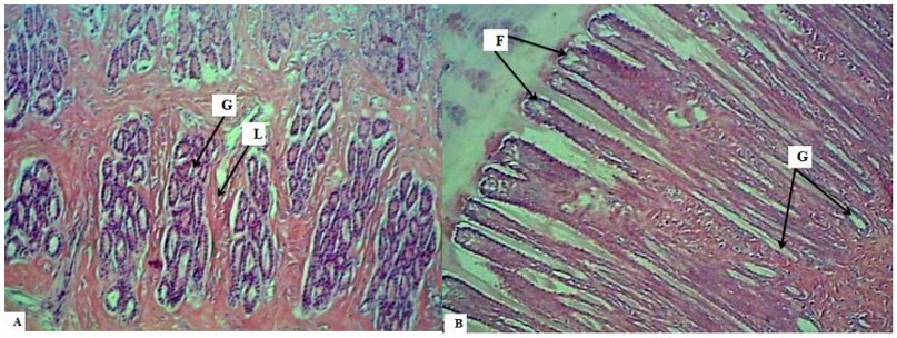
Figure 4. Photomicrograph illustrates the Ventricular showed: Mucosal Foldes(F), Mucosal glands or proprial Glands(G), Connective tissue of Lamina propria(L), A- X 10 H&E in Goose B- X 10 H&E in Guinea Fowl.
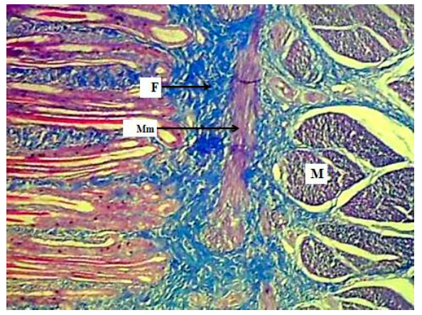
Figure 5. Photomicrograph illustrates the Ventriculas in Male Guinea Fowl showed: collagenous fibers of propria (F), Muscularis mucosa(mM), Muscularis(M) 10X Masson's Trichrome Stain.
A well-developed muscular layer in two studied birds which comprised of fibers of smooth muscles organized in thick inner circular and outer thin longitudinal layers and narrow lines of connective tissue observed between the smooth muscle fibers (Fig.6), The mean thickness of this layer was significantly higher in geese than in guinea fowls (Table1), The serosal layer was formed by loose connective tissue covered by a mesothelium (Fig.7). The mean thickness of this layer was higher none significantly in guinea fowl than in geese (Table 1).
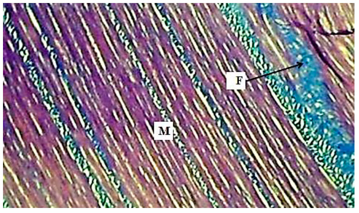
Figure 6. Photomicrograph illustrates the Ventriculas in Male Guinea Fowl: Muscular layer (M), Collagenous fibers (F), 10X Masson's Trichrome Stain.
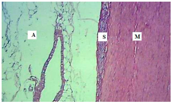
Figure 7. Photomicrograph illustrate the Ventriculas in Male Guinea Fowl showed: Serosal layer ( S), Muscular layer (M), Adipose tissue(A) 10X H&E Stain.
Histochemical
The ventriculus surface epithelium was covered by a layer of Cuticle which gave a pinkish color due to a positive reaction to PAS stain(Fig.8,9). On applying the PAS stain, the secretory material within the lumina and the epithelial cells lining the folds of ventricles positively reacted toward PAS technique(Fig.9A), indicating the presence of neutral mucin. In combination with AB-PAS stain (2.5-PH), the Cuticle covering of ventricular mucosa gave rise to a positive reaction toward this technique (Fig10) due to finding the neutral to acid mucin. A similar response with the same technique (positively) was shown in gastric crypts and the cells of the epithelial lining of ventriculus which indicate to found of these types of mucin (acidic and neutral) (Fig.11,12).
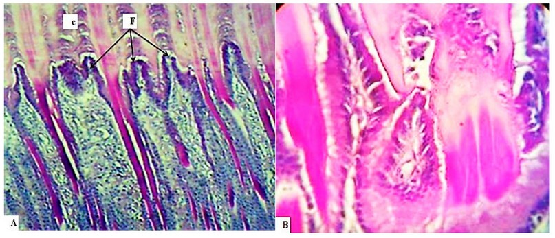
Figure 8. A, B: Photomicrograph illustrate the Ventriculas in Male Goose showed: Cuticle(C), Mucosal Folds(F), Epithelium (E) A-10X PAS Stain. B- 40X PAS
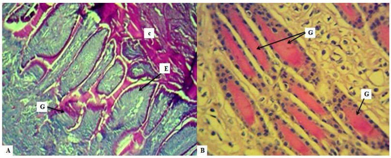
Figure 9. Photomicrograph illustrate the Ventriculas showed: Cuticle(C), Epithelium (E), Folds(F), Glands
A- X 10 PAS Stain. in Guinea Fowl B- Mucosal Glands in Lamina propria 40X PAS in Geese.
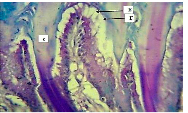
Figure 10. Section illustrate Showed Ventriculas Male Guinea Fowl showed: Cuticle(C), Epithelium (E), Folds(F),
40X PAS-AB Stain.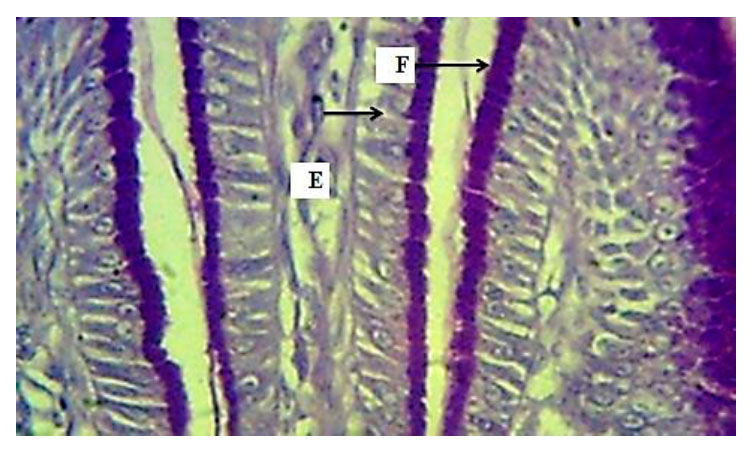
Figure 11. Photomicrograph illustrates Ventriculas in Male showed Epithelium(E), Folds(F), 40X PAS-AB Stain.
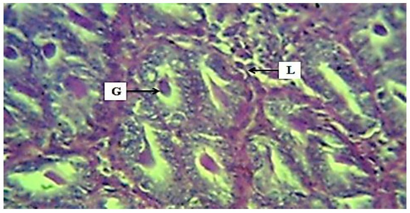
Figure. 12. Photomicrograph illustrates the ventriculus in male Guinea showed: Mucosal crypts or Glands (G), Lamina propria(L) 10X PAS-AB Stain.
DISCUSSION
The microscopic findings observed that the ventricular wall was comprised of three layers, and the submucosa was not present; this result disagrees with 9 in the barn owl. A cuticle covered the mucous membrane, comparable with 10 in pigeon Columba palumbus. The mean thickness of this layer was significantly higher in geese than in guinea fowls. The absence or presence and thickness of Cuticle were related to the type of the bird's diet 11. The thin Cuticle was found in frugivores while a thick Cuticle was observed in granivores;, a layer of cuticle act like a crushing surface which facilitates digestion of food mechanically by the muscular stomach, therefore the granivorous, insectivorous and herbivorous own a thick layer of the Cuticle as well as the firm stomach was distinctively developed12. The mucosa was thrown into longitudinal mucosal folds lining with mostly simple cuboidal to columnar cells as found in blue and yellow macaws by 13. The proprial glands (tubular mucous glands) are lined by simple cuboidal epithelium, a similar result recorded in Mallard by14.
The mean height of lining epithelial cells was higher non significantly. Still, the mean length of folds and mean thickness of tunica mucosa were substantially higher in geese than in guinea fowl. The propria was filled by gastric glands (simple tubular glands), which were arranged into two shapes; the first appeared long and straight, while the second present as short, oval-shaped and packed; a similar result was reported by 6; also, the propria was rich by the collagenous fibers, while the muscular mucosa appeared interrupted. This finding was parallel to the findings recorded by15 in Brown Falcon but disagreed with Black-tailed Crake by 16.
The submucosal layer was absent; this was agreed with17 in coot birds but disagreed with 18 in black-winged Kite. A well-developed muscularis in two studied birds comprised outer thin longitudinal and inner thick circular layers of smooth muscle fibers. The mean thickness of this layer was significantly higher in geese than in guinea fowl, this observation was following that found by 18, but 13 noticed only one developed circular layer was present in Blue and Yellow macaws, while 15 reported in Brown Falcon this layer was constructed from three orientations; thin inner and outer longitudinal, and in between thick circular. Development of this layer may concern the bird's mechanical grinding capability to the ingested foods; the contracted ventricular muscular act to mechanically digest the consumed foods; thus, the avian herbivorous, insectivorous, and granivorous have a thick cuticle layer and muscular ventriculus. In contrast, species that eat food that unrequired mechanical digestion significantly as piscivores or carnivores possesses mostly a thin or undeveloped ventriculus with a thin cuticle 12, 14. The serosal layer was formed by loose connective tissue covered by a mesothelium; this finding was concerned with previously observed in 9 in mallard and Columba palumbus. The mean thickness of this layer was higher (non significantly) in guinea fowl than in geese.
CONCLUSION
The study concluded that presence of similarity in histological composition in ventriculus of two birds (goose and guinea fowls) with some significant micro morphometric measurements differences and similar to those reported in herbivorous, granivorous and insectivorous species which have thick Cuticle and ventriculus, but differ from of carnivores and piscivores which possesses a rudimentary ventriculus and soft Cuticle that not require significant mechanical digestion and this differences may be due to the adaptation to the food types.
Acknowledgment: Thanks going for all who support us.
The conflict between authors: No conflict
Funds: self by authors
REFERENCES
1. Clench MH, Mathias JR. The avian cecum: a review. The Wilson Bulletin. 1995:93-121.
2. Ahmed YA, Kamel G, Ahmad AA. Histomorphological studies on the stomach of the Japanese quail. Asian Journal of Poultry Science. 2011;5(2):56-67.
3. Moţ M. Morphological aspects of digestive apparatus in owl (Asio flammeus) and dove (Columba livia). Lucrari Stinifice MedicinaVeterinara. 2010;8(2):364-7.
4. Taylor M. Anatomy and physiology of the gastrointestinal tract for the avian practitioner. Birds. Post Grad Found in Vet. Sci. Uni. of Sydney, Aust. Proc. 2000;334:107-13.
5. Al-Saffar FJ. Histomorphological and histochemical study of stomach of domestic pigeon (Columba livia domestica): FJ Al-Saffar1 and Eyhab, RM Al-Samawy2. The Iraqi Journal of Veterinary Medicine. 2016;40(1):89-96.
6.Abumandour MM. Morphological studies of the stomach of falcon. Scientific Journal of Veterinary Advances. 2013;2(3):30-40.
7. Bancroft JD, Stevens A, Turner R. Theory and practice of histological techniques. 2nd (ed.) churchill living stone. New York. 1982.
8. Thiem A, Dusa A. Qualitative comparative analysis with R: A user's guide. Springer Science & Business Media; 2012.
9- Al-Juboory RW, Dauod HA, Al-arajy AS. Comparative anatomical and histological study of the stomach in two Iraqi birds (Columba palumbus and Tyto alba). Ibn AL-Haitham Journal For Pure and Applied Science. 2017;29(2):1-2.
10. Al-Juboury RW. Comparative anatomical and histological study on the digestive tract in two Iraqi birds, common wood pigeon Colimba palumbus (L.) and barn owl Tyto alba (Scopoli). Pure. Appl. Sci. 2016;24(5):946-56.
11. Bailey TA, Mensah-Brown EP, Samour JH, Naldo J, Lawrence P, Garner A. Comparative morphology of the alimentary tract and its glandular derivatives of captive bustards. The Journal of Anatomy. 1997;191(3):387-98.
12. Denbow DM. Gastrointestinal anatomy and physiology in sturkies avian development. British Poultry Science. 2000;42:505-13.
13Rodrigues MN, Abreu JA, Tivane C, Wagner PG, Campos DB, Guerra RR, Rici RE, Miglino MA. Microscopical study of the digestive tract of Blue and Yellow macaws. Current microscopy contributions to advances in science and technology (A. Méndez-Vilas, Ed.). 2012:414-21.
14. Al-Samawy ER. Histomorphological and histochemical comparsion of the stomach and small intestine of the domestic pigeon (Columba Livia domestica), Striated Scope Owl (OtusScorsbrucei) and mallard (Anasplatyrhynchos)[Thesis]. Baghdad, Iraq: University of Baghdad. 2015.
15 Al-Taee AA. Macroscopic and microscopic study of digestive tract of brown falcon Falco berigora in Iraq. JUBPAS. 2017;25(3):915-36.
16. Zhu L. Histological and histochemical study on the stomach (proventriculus and Gizzard) of black-tailed crake (Porzana bicolor). Pakistan Journal of Zoology. 2015;47(3).
17. Batah AL, Selman HA. Histological study for stomach (proventriculus and Gizzard) of coot bird Fulica atra. Diyala Journal of Agricultural Sciences. 2012;4(1).
18. Hamdi H, El-Ghareeb A, Zaher M, Essa A, Lahsik S. Anatomical, histological and histochemical adaptations of the reptilian alimentary canal to their food habits: II-Chamaeleon africanus. World Applied Sciences Journal. 2014;30(10):1306-16.
Received: 5 January 2022 / Accepted: 27 July 2022 / Published:15 August 2022
Citation: Khaleel I M, Abd Alkhazraji K I, Senan Hasan M. A Comparative Histomorphological and Histochemical Study of the Ventriculus between Iraqi Adult Geese (Anser anser) and Guinea fowls (Numidia meleagris). Revis Bionatura 2022;7(3) 37. http://dx.doi.org/10.21931/RB/2022.07.03.37