2023.08.03.96
Files > Volume 8 > Vol 8 No 3 2023
Histological changes and diagnostic value of immunoglobulin G/M to Helicobacter pylori in gastric cancer patients

Ahmed Nassir Faisal 1* , Raed Madhi2, Anwar Algaber3
1 National university of science and technology, Thi-Qar 64001, Iraq.
2Department of Biology, Collage of Science, Misan University, Maysan 62001, Iraq.
3Mazaya University College, Thi-Qar 64001, Iraq.
* Corresponding author [email protected]
Available from: http://dx.doi.org/10.21931/RB/2023.08.03.96
ABSTRACT
Stomach cancer is believed to be one of the most common cancers that lead to death. In Iraq, stomach cancer occupies the seventh place of cancer occurrence in both sexes and is counted as one of the ten most common cancers.
The current study is designed to explore the link between Helicobacter pylori (H.pylori) infection and the development of incidences of stomach cancer. In addition, related age and gender were also studied. Histological examinations of stomach biopsies were performed in suspected people to evaluate stomach cancer occurrences. Of the 40 patients with stomach cancer, the infection of H. pylori was emphasized in 34 (66.66%) with serum IgG/IgM, which reflected a significant frequency for infection of H. pylori in stomach cancer patients. The study also showed that males with H. pylori infection record a higher percentage than female patients with stomach cancer. Moreover, the results revealed that age is also connected to H. pylori infection. Based on the above findings, monitoring infected people with H. pylori might be an excellent strategy to control stomach cancer occurrences.
Keywords: Stomach cancer, Infectious diseases, IgG, IgM, H. pylori.
INTRODUCTION
Stomach cancer, in general, is considered one of the world's most significant public health problems and the most prominent challenges facing the scientific community in the current century due to the high rates of infection and mortality of cancer. Stomach cancer is one of the most common cancers that can lead to death and is also shown to be connected with cancer worldwide1,2. The development of this type of cancer is linked to several risk factors, and the incidence of this disease lags by age, socioeconomic conditions, and geographical location3. Indeed, stomach cancer is ranked seventh with 3.67% among all other body cancers. Carcinoma of the gastric adenocarcinoma accounts for 90–95% of all other gastric carcinomas, and in Lorraine 1965, gastric carcinomas can progress to the intestinal type4.
Stomach cancer is the fifth most common, after lung, colon, and prostate cancer. It is the third cancer leading to deaths worldwide, at the end of 782,685 cases, or 8.2%. With the changes in the International Agency for Research on Cancer of the World Health Organization (World Health Organization WHO Soba), nearly one million good cases of stomach cancer are recorded5 annually. Gastric adenocarcinoma of the epithelial area is 90-95% of other stomach cancer6,7.
Many environmental and genetic factors are linked to stomach cancer development, all of which can play a role in stomach cancer8. For instance, cigarette smoking and drug abuse9,10, Nutritional factors11,12, Obesity13, and occupational exposure14. Moreover, other potential risk factors that might increase the risk of stomach cancer incidences are gastric surgery, gastroesophageal reflux disease, physical inactivity, non-steroidal anti-inflammatory drugs (NAIDs) and exposure to radiation15-17. The infection of H. pylori might be a risk factor connected to stomach cancer development. In 1994, the International Agency for Research on Cancer classified H. pylori among the causes of cancer of the class or first degree (Class 1 carcinogen). H. pylori is a gram-negative, air-loving, motile bacterium with 4-6 flagella helping penetrate the gastric mucosa of both the stomach and duodenum18. H. pylori infection rates increase with age due to the increasing cumulative effect, and the development of stomach cancer in those people might be connected to H. pylori infection19-22. It has been established that infection with H. pylori is accompanied by a decrease in hydrochloric acid (HCI) secreted by parietal cells, creating a suitable environment for survival. After H. pylori enters the stomach cavity, it secretes the urease enzyme that divides the urea compound into ammonia (NH3) and carbon dioxide (CO2)23. These components provide an alkaline environment, which leads to surrounding them with an alkaline medium equivalent to gastric acidity. Accordingly, they are not affected by stomach acid like other types of bacteria; in addition to this enzyme, they produce many different enzymes, such as catalase enzyme24.
H. pylori infection and taking (NAIDs) are the most common cause of gastric ulcers. Gastric ulcers occur in the surface epithelium, extend to the mucous layer in the stomach wall, and, over time, can infect the peritoneum. Several studies have documented that people with gastric ulcers and duodenal ulcers have a higher risk of developing stomach cancer25. Therefore, monitoring people with H. pylori infection might pave the way to control stomach cancer.
MATERIALS AND METHODS
Study design
The current study was performed in the Thi-Qar province/ southern Iraq. The ethical approval for this study was obtained from the Al-Nassrya Health Department.
The present study included 51 patients with stomach cancer who had undergone total or partial gastrectomy at the Cancer Center of Al-Hussein Teaching Hospital of the Dhi-Qar Health Department. Diagnosis of stomach cancer was performed by the specialist medical staff at Al-Hussein Teaching Hospital based on clinical examinations and stomach biopsy. Furthermore, serum IgG/IgM sensitivity was performed to detect H. pylori infection.
Histological examinations
Diagnosis of stomach cancer was done by taking total or partial gastrectomy of suspected people with stomach cancer. (4%) formaldehyde fixed tissue samples were subjected to serial dehydration and paraffin embedding. Then, (H&E) stained samples were evaluated under light microscopy. The evaluations of stomach cancer grading were done according to standard protocol 26.
Estimation of IgG/IgM sensitivity of H. pylori
H.pylori was identified using the H. pylori antibody (IgG/IgM) diagnostic Kit (RC11, ANAMOL, Kolgaon) and based on the manufacturer's instructions. The test line was provided with anti-IgG and anti-IgM, and the purple color on the test line was used to indicate positive H. pylori.
Statistical analysis
Statistical tests were done using the Chi-square test, and data were introduced as a percentage. P < 0.05 is considered statistically significant between the groups.
RESULTS
Histological study
For stomach cancer diagnosis, a stomach biopsy was carried out in suspected people with stomach cancer. Histological examinations showed a clear-cut destruction in the architectures of harvested tissue samples. Variations in the size and shape of the malignant cells characterized these destructions. For example, the histological results showed pleomorphic irregular cells and their infiltration and penetration to different degrees within the layers of the stomach wall, as shown in (Figure). As for the histological pattern, the results of the current study show that epithelial adenocarcinoma is the most common type.
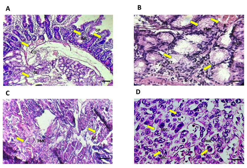
Figure 1. Cross-section of gastric cancer tissue using hematoxylin & eosin. Scale bar = 50 μm. A) changes in the lumen of glandular structures (GL) with cases of congestion (CO) and extensive infiltration of lymphocytes (LC) In gastric pits (GP). B) hemorrhage (B) with inflammatory cell infiltration (INF) in the Fundus region with atrophy and disappearance of gastric glands (AT.). C) glandular cavities )GC) and invasion of the muscular layer by polymorphous carcinomas (PMC). D) the invasion of polymorphous tumor cells (PMC) represented by signet ring cells (SRC) with terminal site nucleus and dense chromatin(N).
Age group
In the present study, we found that the age group (≥ 50 years old) recorded a remarkable increase in H.pylori infection of patients with stomach cancer, which represented 79.41% (Figure 4), compared to the age group (30-39 and 40-49) that correspond to the percentages 11.76% and 8.83%, respectively (Figure 2).
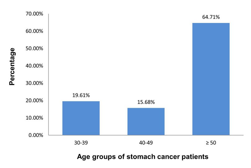
Figure 2. Age groups of patients with stomach cancer. Age criteria were divided into three groups (30-39, 40-49, ≥ 50). Data were presented as a percentage.
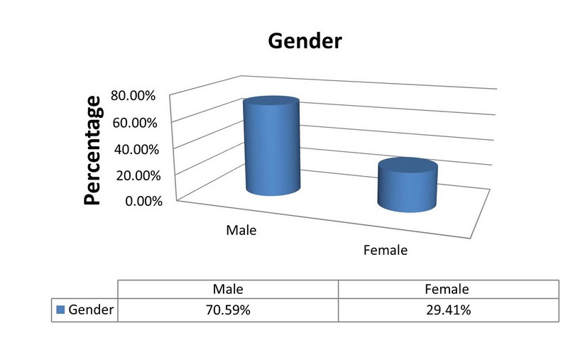
Figure 3. Distribution of H. pylori infection among males and females of stomach cancer patients. Data were introduced as a percentage. The chi-square test was used for comparison between the groups. The significant difference between the groups was considered as P-value £ 0.05.
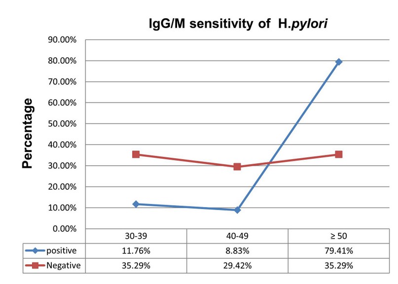
Figure 4. Percentages of IgG/M of H. pylori infection between age groups of stomach cancer patients. The blue line is the positive infection of H. pylori. The red line indicates a harmful infection of H. pylori. Data were presented as a percentage. The chi-square test was used for comparison between the groups. The significant difference between the groups was considered as P-value £ 0.05.
Gender
The results showed a remarkable superiority of males over females in the samples of stomach cancer patients infected with H. pylori. The frequency of the infection of H. pylori increased significantly (p < .05) in males with stomach cancer, compared to females with stomach cancer and corresponding to the percentages 70.59% and 29.41%, respectively, as shown in (Figure 3).
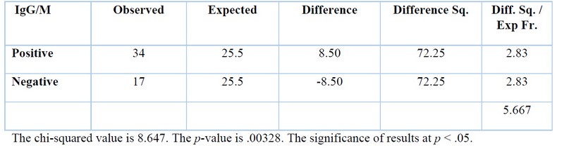
Table 1. Statistical differences between male and female patients with stomach cancer.
IgG/M detection of H.pylori infection
In this study, the sensitivity of IgG/M to the infection of H. pylori in patients with stomach cancer was examined using the sensitivity to IgG/M (Figure 4 and Table 2). We found that the frequency of infection of H. pylori was significantly higher in the IgG/M positive group compared to the IgG/M negative group (Table 2). It is essential to mention here that the infection of H. pylori recorded the highest percentage (76.41%) in the age group (≥ 50) of patients with stomach cancer as compared with another age group (Figure 4).
Table 2. Statistical differences between IgG/M positive and negative of patients with stomach cancer.
DISCUSSION
It is well established that stomach cancer is the most common cancer and one of the leading causes of death worldwide, particularly in developing countries1,2. The possible incidence of stomach cancer can be due to different risk factors. In the current study, our findings showed that the infection of H. pylori significantly correlates to stomach cancer incidences. Moreover, the results also found a significant link between age and gender and H. pylori infection. Thus, detecting people with H. pylori infection could be an excellent way to control stomach cancer occurrences.
Various risk factors have been demonstrated to be connected to the development of stomach cancer9,11,27. It has been shown that the age factor is significantly associated with infection of H.pylori and the product of stomach cancer28. In the current study, we found that H.pylori infection was higher (79.41% ) in the age group (≥ 50) of patients with stomach cancer. Our results aligned with accumulating data showing that age factor is significantly associated with H. pylori infection28. A recent study has explored that H. pylori infection plays an essential role in the development of stomach cancer29, which could explain the risk of age factor to stomach cancer incidences.
Gender is another factor suspected to have a link with H. pylori infection participating in the development of stomach cancer30; thus, in the present study, we examined the correlation between gender and the condition of H. pylori. The current data showed that males with stomach cancer recorded a higher percentage (70.59% ) of H. pylori infection than females (Figure 3). Our data aligned with a recent study showing that the favorable ratio of H. pylori infection was significantly higher in the male group (43.68%) than in the female group (28.8%). Thus, periodic examinations for males could help avoid the infection with H. pylori, which might also protect against stomach cancer incidences.
It is well known that infection of H. pylori could be developed and cause chronic inflammation, which can increase the possibility of stomach cancer occurrences31. Moreover, H. pylori infection has also been considered the most common risk factor for stomach cancer26,30,32. We showed that the sensitivity of IgG/M to the disease H. pylori in patients with stomach cancer was higher in the IgG/M positive group than in the IgG/M negative group (Figure 4 and Table 2). The results also demonstrated that the infection of H. pylori was significantly increased (76.41%) in the age group (≥ 50) of patients with stomach cancer as compared with another age group (Figure 4). Therefore, this could explain why the infection with H. pylori might extensively participate in stomach cancer incidence connected with age factor. The exact mechanism of H. pylori-induced stomach cancer incidence is not entirely understood. However, the explanation of this condition could be due to the penetration of H. pylori to the mucous layer by flagella; scratching results in stimulating host cells to produce mediators of inflammation such as increased tumor necrosis factor (TNFα) and interleukins that promote the attraction of inflammatory cells22. These bacteria also have many strains that differ in virulence factors as a result of differences in two types of genes; the first is suspected to be responsible for the incidence of stomach cancer and associated with cytotoxicity (Cytotoxin associated gene) (CagA) ، while the other gene is the vacuolating cytotoxicity gene (Vaculating cytotoxin gene) (Vag A), which is responsible for creating vacuoles and perforations in the stomach tissue, which result in the activation of programmed death of stomach epithelial cells30,33.
CONCLUSION
The present data demonstrate that age and gender strongly correlate to the infection of H.pylori that participates in the incidences of stomach cancer. Furthermore, our findings also show that the condition of H.pylori is extensively involved in stomach cancer occurrences. Accordingly, infection of H.pylori could have a high risk of stomach cancer incidences, and therefore, monitoring people with H.pylori infection might control the development of stomach cancer.
Acknowledgment
We are thankful to the Al-Nassrya Health Department for providing us with authorization approval for the current study. We are also grateful to the National University for Science and Technology College of Health and Medical Technology, Mazaya University College and the University of Misan for their support during this study.
Conflict of interest: The authors announced that the present study has no conflict of interest.
REFERENCES
1- Ilic, M. and I. Ilic, Epidemiology of stomach cancer. World J Gastroenterol, 2022. 28(12): p. 1187-1203.
2- Figueiredo, C.; Camargo, M.; Leite, M.; Fuentes-Pananá, E.; Rabkin C. and Machado, J. C. (2017). Pathogenesis of gastric cancer: genetics and molecular classification. Microbiol. Immunol., 400:277-304.
3- ICR (Iraqi Cancer Registry). (2016). Annual Report. Ministry of Health, Baghdad-Iraq: I+ 287pp.
4- Shah, S. C., McKinley, M., Gupta, S., Peek Jr, R. M., Martinez, M. E., & Gomez, S. L. (2020). Population-based analysis of differences in gastric cancer incidence among races and ethnicities in individuals age 50 years and older. Gastroenterology., 159(5): 1705-1714.
5- Farhood, B., G. Geraily, and A. Alizadeh, Incidence and Mortality of Various Cancers in Iran and Compare to Other Countries: A Review Article. Iran J Public Health, 2018. 47(3): p. 309-316.
6- Carvalho, C. E.; McCormick, T. M.; Carvalho, P. C.; Fischer, J. S.; Aquino, P. F. ; Neto, G. P. B. and Carvalho, M. D. (2016). Considerations about gastric cancer proteomics. Rev. Col. Bras. Cir., 43(5): 395-397.
7- Awad, H. A.; Hajeer, M. H.; Abulihya, M. W.; Al-Chalabi, M. A.; Al Khader, A. A. (2017). Epidemiologic characteristics of gastric malignancies among Jordan University Hospital patients. Saudi Med. J., 38(9): 965-967.
8- Yu, S.; Yu, Y.; Zhao, N.; Cui, J.; Li, W. and Liu, T. (2013). C-Met as a prognostic marker in gastric cancer: A systematic review and meta-analysis. PLoS ONE, 8(11): 79137-79137.
9- Cheng, X. J.; Lin, J. C. and Tu, S. P. (2016). Etiology and prevention of gastric cancer. Gastrointestinal Tumors., 3(1):25–36.
10- Karimi, P.; Islami, F.; Anandasabapathy, S.; Freedman, N. D. and Kamangar, F. (2014). Gastric cancer: Descriptive epidemiology, risk factors, screening, and prevention. Cancer. Epidemiol. Biomar. Prev., 23(5):700-713.
11- Sitarz, R.; Skierucha, M.; Mielko, J.; Offerhaus, G. J. A.; Maciejewski,R. and Polkowski, W. P. (2018). Gastric cancer: Epidemiology, prevention, classification, and treatment. Cancer Man. and Res., 10: 239–248.
12- Danyal, M.; Ahmad, S.; Ahmad, M.; Asif, H, M.; Akram, M.; Ur Rehman, S. and Sultana, S. (2015). Risk factors and epidemiology of gastric cancer in Pakistan. Asian, Pac. J. Cancer Prev., 16(12):4821-4824.
13- Fan, J. h ; Wang, J. B.; Wang, S,H ‐M.; Abnet, C,H. C.; Qiao, Y.; and Taylor, P. R. (2017). Body mass index and risk of gastric cancer: A 30‐year follow-up study in the Linxian general population trial cohort. Can. Sci., 108(8): 1667–1672.
14- Santibanez, M.; Alguacil, J.; de lahera, M. G.; Navarrete-Munoz, E. M.; Llorca, J.; Aragones, N.; Kauppinen, T. and Vioque, J. (2012). Occupational exposures and risk of stomach cancer by histological type. Occup. Environ. Med., 69(4):268-275.
15- Lee, Y. Y.; Lee, and Derakhshan, M. H. (2013). Environmental and lifestyle risk factors of gastric cancer. Arch. Iran Med., 16(6): .365 – 358.
16- Kong, P.; Wu, R.; Liu, X.; Liu, J.; Chen, S,H ; Ye, M.; Yang, Ch.; Song, Z.; He, W.; , Yin, Ch.; Yang, Q.; Jiang, Ch.; Liao, F.; Peng, R.; Zhou, Z.; Xu, D. and Xia, L. (2016). The effects of anti-inflammatory drug treatment in gastric cancer prevention: an update of a meta-analysis. J. Cancer, 7(15): 2247-2257.
17- Kim, S. M.; Min, B. H. ; Lee, J.; An, J. Y.; Lee, J. H; Sohn, T. S.; Bae, J. M.; Kim, J. J.; Kang, W. K.; Kim, S. and Choi, M-G. (2018). Protective effects of female reproductive factors on Lauren intestinal-type gastric adenocarcinoma. Yonsei. Med. J., 59(1):28-34.
18- Marshall, B. J. and Warren, J. R. (1984). Unidentified curved bacilli in the stomach of patients with gastritis and peptic ulceration. Lancet, 1(8390): 1311–1315.
19- Lazar. D. (2013). Gastric carcinoma new insights into current management. Intech Open, London: 1-293pp.
20- Kirshners, A.; Polaka, I. and Aleksejeva, L. (2015). Gastric cancer risk analysis in unhealthy habits data with classification algorithms. Infor. Technol. Man. Sci., 18(1): 97-102.
21- Alsamarai, A. M.; Alsaadi, F. T,H M.; Mohamed, F. I.; Alobaidi, A. H, A. and Alhili, M. B. (2018). Helicobacter Pylori strains isolated from Iraqi subjects with gastritis, peptic ulcer, and carcinoma of stomach genetic sequences: A new variants. Adv. Res. Gastroenterol. Hepatol., 11(5): 1-8.
22- Alsamarai, A. M.; Alsaadi, F. T,H M.; Mohamed, F. I.; Alobaidi, A. H, A. and Alhili, M. B. (2018). Helicobacter Pylori strains isolated from Iraqi subjects with gastritis, peptic ulcer, and carcinoma of stomach genetic sequences: A new variants. Adv. Res. Gastroenterol. Hepatol., 11(5): 1-8.
23- Stewart, B. W. and Wild, C. (2014). World Cancer Report 2014. International Agency for Research on Cancer, and World Health Organization. Lyon, France, Geneva, Switzerland. http://publications.iarc.fr.
24- Piazuelo, M. B. and Correa, P. (2013). Gastric cancer: Overview. Colombia Med., 44(3): 192-201.
25- Kumar, V.; Abbas, A. K. and Aster, C. (2013). I am running primary pathology. 9th ed. Elsevier, Book aid and Sabre foundation, Canada: VIII+910pp.
26- Hu, B., et al., Gastric cancer: Classification, histology and application of molecular pathology. J Gastrointest Oncol, 2012. 3(3): p. 251-61.
27- Boccolini, P. M. M.; Asmus, C. I. R.; Chrisman, J. R.; Câmara, V. M.; Markowitz, S. B. and Meyer, A. (2014). Stomach cancer mortality among agricultural workers: Results from a death certificate-based case-control study. Cad. Saúde Colet., Rio de Janeiro, 22 (1): 86-92.
28- Huang, Q., et al. (2021). Infection in Geriatric Patients: Current Situation and Treatment Regimens. Front Med (Lausanne). 8: p. 713908.
29- Díaz, P., et al. (2018). Gastric Cancer: Adaptive Cellular Mechanisms Involved in Disease Progression. Front Microbiol, 9: p. 5.
30- Zhao, X.X., et al. (2020). Effect of Gender and Age on the Correlation between Helicobacter pylori and Colorectal Adenomatous Polyps in a Chinese Urban Population: A Single Center Study. Gastroenterol Res Pract, 2020: p. 8596038.
31- Rivas-Ortiz, C. I.; Lopez-Vidal, Y.; Arredondo-Hernandez, L. J. R. and Castillo-Rojas, G. (2017). Genetic alterations in gastric cancer associated with Helicobacter pylori infection. Fron. Med.,4(47)1-12.
32- Muzaheed. (2020). Oncogenicity: Mechanism, Prevention, and Risk Factors. ScientificWorldJournal, 2020: p. 3018326.
33- Queiroz, D. M. M.; Harris, P. R.; Sanderson, I. R.; Windle, H. J.; Walker, M. M.; Rocha, A. M. C.; Rocha, G. A.; Carvalho, S. D.; Bittencourt, P. F. S.; de Castro L. P. F.; Villagra ́n, A.; Serrano, C.; Kelleher, D.; Crabtree, J. E. (2013). Iron status and Helicobacter pylori infection in symptomatic children: An international multi-centered study. PLOS ONE, 8(7): 68833- 68838.
Received: 25 June 2023/ Accepted: 26 August 2023 / Published:15 September 2023
Citation: Nassir Faisal A, Madh R, Algabe A. Histological changes and diagnostic value of immunoglobulin G/M to Helicobacter pylori in gastric cancer patients. Revis Bionatura 2023;8 (3) 96 http://dx.doi.org/10.21931/RB/2023.08.03.96