2023.08.01.46
Files > Volume 8 > Vol 8 No 1 2023
Molecular analysis of Fungi: Malasseziarestricta from Felidae

Afkar Muslim Hadi*¹ , Hani Saber Khalif ²
¹Iraq Natural History Research Center and Museum, University of Baghdad;
²Iraq Natural History Research Center and Museum, University of Baghdad; email: [email protected]
*Correspondence: e-mail:[email protected]
Available from: http://dx.doi.org/10.21931/RB/2023.08.01.46
ABSTRACT
A total of 9 samples of wild cat Felischausfurax (de Winton, 1898) and 13 (11 positives) samples of domestic cat Feliscattus (Linnaeus, 1758) belong to Family Felidae. All cats were trapped and used hair and skin scrapings by forceps and surgical blades. The areas of the collection were: Mosul province (north of Iraq); Baghdad, Al-Rashidiya, Tharthar, Nahrawan, AL-Mahmoudiya (middle of Iraq) and AL-Haretha (south of Iraq). The current study revealed that the sensitive and specific PCR assay allowing rapid and reliable identification of Malasseziarestricta by the fragment size amplified was 500bp in the ITS1 gene in one sample of wild cats. The current study recorded a new strain of Malasseziarestrictathat called AF2013 strain "small subunit ribosomal RNA gene, partial sequence; internal transcribed spacer 1", complete sequence; and 5.8S ribosomal RNA gene, partial sequence. Which was inserted in GenBank: MW376484.1 from wild cat Felischausfurax for the first time in Iraq. Sequencing revealed close matching of the phylogenetic tree to an isolate from Korea (CP030254). The compression was performed using NCBI – the based nucleotides website.
Keywords: Dermatitis, Cutaneous microflora, fungi, Genotype, Malasseziarestricta.
INTRODUCTION
The taxonomy of Malasseziarestricta(E.Guého, J.Guillot&Midgley) according to GBIF 1:
Kingdom: Fungi
Phylum: Basidiomycota
Class: Malasseziomycetes
Order: Malassezialess
Family: Malasseziaceae
Genus: MalasseziaBaill, (1889)
Synonyms: PityrosporumSabour (1904)
The genus of Malassezia includes seven species comprising the three former taxa M. furfur, M. pachydermatis and M.sympodialis, and four new taxa M. globosa, M. obtuse, M. restricta and M. slooffiae. 2.
Molecular techniques have increased research in this field, such that revealed the infection with Malassezia spp. to man for over 150 years, and seven new species have been added to this genus 3.
The skin colonization with Malassezia spp. depends on the skin's body site, the host's age, and other comorbid skin conditions, as the geographic area. It is found in the highest density in sebaceous regions such as the scalp, face, and upper trunk. It is seen in higher densities in young adults, who tend to have relatively oily skin, 4.
Malassezia spp. is naturally found on the skin surfaces of many animals, including humans. It can cause hypopigmentation or hyperpigmentation on the trunk and other locations in humans, 5. The knowledge about Malassezia spp. has expanded remarkably since the 1990s in dogs and cats, 6 . Malassezia furfurwas isolated from cats in 1999 by Crespo 7.
The epidemiological map of the spread of fungi Malassezia from animals to humans is unclear, as Morris 8 indicated that M. pachydermatis was transmitted from pet dogs to humans. This is an important indicator that prompts us to research further the transmission of Malassezia spp. from domestic and wild animals. In Poland, along with surveys from 2008 to 2018 for many groups of animals like dogs, cats, rodents, riding horses, birds and other pet animals (reptiles and mammals), Bozena 9 revealed to fungal species involved and evaluated the risk of their transmission to humans.
The study aims to spotlight the species of Malasseziarestrictathat infected domestic and wild cats through molecular analysis for the first time in Iraq.
MATERIALS AND METHODS
Collection of samples: A total of 9 samples of wild cat Felischausfurax (de Winton, 1898) and 13 (11 positives) samples of domestic cat Feliscattus (Linnaeus, 1758 ) ofFelidae Family were trapped and used as hair and skin scrapings by forceps and surgical blades. The scales were collected in sterile empty Petri dishes10The areas of the collection were: Mosul province (north of Iraq); Baghdad, Al-Rashidiya, Tharthar, Nahrawan, AL-Mahmoudiya (middle of Iraq) and AL-Haretha (south of Iraq).
DNA Extraction
Genomic DNA was isolated from two samples according to the QIAamp DNA Mini Kit protocol, QIAGEN.
Primer preparation
Lyophilized primers (Macrogen Company) were dissolved in nuclease-free water to give a final concentration of 100 pmol/μl as a stock solution. A working solution of these primers was prepared by adding 10μl of primer stock solution (stored at freezer -20 C) to 90μl of nuclease-free water to obtain a working primer solution of 10 pmol/μl. Tables 1, 2 and 3.
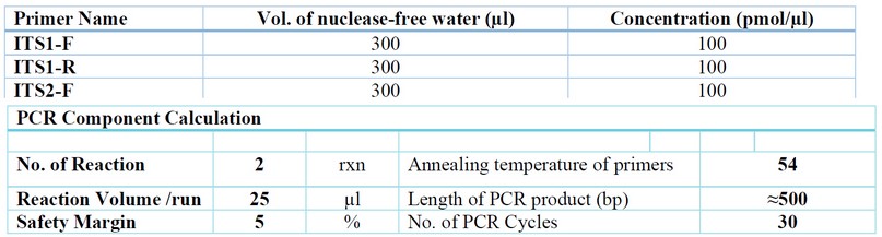
Table 1. Primer preparation as Macrogen Company protocol.
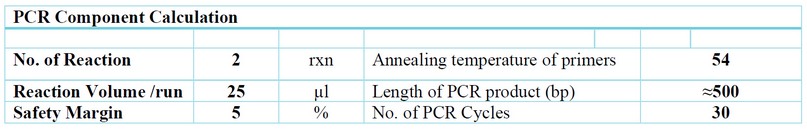
Table 2.Reaction Setup and Thermal Cycling Protocol.
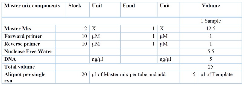
Table 3. PCR Program.
Agarose Gel Electrophoresis was adopted to confirm the presence of PCR amplification.PCR products were loaded directly. The Ethidium bromide-stained bands in gel were visualized using a Gel imaging system. Standard Sequencing APPLIED IN KOREA (MACROGEN CORPORATION) BY SANGER USING (ABI3730XL).
RESULTS AND DISCUSSION
Summary of Data Production: the results of DNA concentration for two samples were concluded in table 4.
PCR amplification of two samples, ITS1 and ITS2, as in Figures 1 and 2.
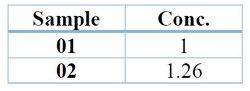
Table 4. DNA Concentration (ng/µl).
The current study revealed that the sensitive and specific PCR assay allowing rapid and reliable identification of Malasseziarestricta by the fragment size amplified was 500bp in ITS1 gene in one sample of the wild cat as in Figure 1. And there is no results shown in the ITS2 gene as in figure 2.
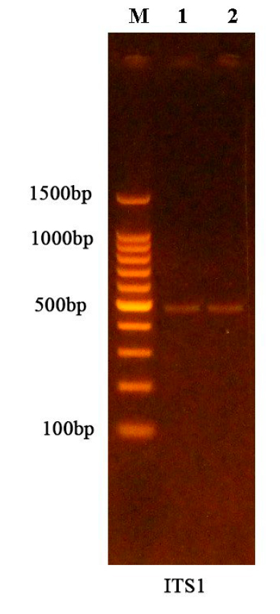
Figure 1. Results of the amplification of ITS1 gene were fractionated on 1.5% agarose gel electrophoresis stained with Eth.Br. M: 100bp ladder marker. Lanes 1-2 resemble ≈500bp PCR products.
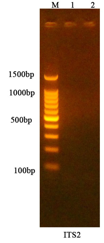
Figure 2. No Results of the amplification of the ITS2 gene were fractionated on 1.5% agarose gel electrophoresis stained with Eth. Br. M: 100bp ladder marker.
Data Analysis
The current study recorded a new strain of Malasseziarestrictathat called AF2013 strain "small subunit ribosomal RNA gene, partial sequence; internal transcribed spacer 1", complete sequence; and 5.8S ribosomal RNA gene, partial sequence, which inserted in GenBank: MW376484.1 from wild cat Felischausfuraxfor the first time in Iraq—fig. 3.
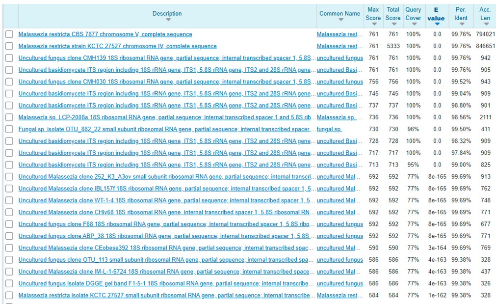
Figure 3. BLAST 2 results of sequences revealed to Malasseziarestricta from wild cat in Iraq.
The current result of recording Malasseziarestricta cat indicates that it is a common disease between humans and animals, where it was found by Sugita 11 revealed that M. restrict commonly colonizes both AD (Atopic Dermatitis) patients and healthy subjects. And more, Annabelle 12 described the case of a pediatric oncology patient with splenic lesions secondary to Malasseziarestricta. Males and females are affected by the infection of (PV) PityriasisVersicolor, especially individuals between (10-20) years, 13. No significant correlation was reported between economic status; type of job; or water source with the infection of Malassezia spp. 14, which increases the risk.
Awad 14 revealed that patients who have contact with dogs only were equally exposed to Malassezia spp., which reflects that dogs are not only the source of infection. Bond 6 reviewed 18 types of fungus Malassezia and their locations between animals and humans and mentioned Malasseziarestricta only in humans.
Phylogenetic tree
The current sequencing data has reportedMalasseziarestricta from Iraq (MW376484); their host is a wild cat; this species revealed close matching on the phylogenetic tree to an isolate of Malasseziarestricta strain KCTC 27527 chromosome IV, complete sequence from Korea (CP030254); their host is Homo sapiens. On the other hand, it matches the isolation of Malasseziarestricta strain Y. H. Yeh I0610 "small subunit ribosomal RNA gene, partial sequence; internal transcribed spacer 1", "5.8S ribosomal RNA gene and internal transcribed spacer 2 , the complete sequence" and large subunit ribosomal RNA gene, partial sequence from Taiwan (MK336446) their host is a goat (Ipomoea pes-caprae). Then, it matches the isolation of Malasseziarestricta CBS 7877 chromosome V, complete sequence from the United Kingdom: Bristol (CP033152); their host is Homo sapiens. Fig. 4.13revealed that the evidence shown by the phylogenetic tree showed that all species of Malasseziaspp. have high similarity with each other.
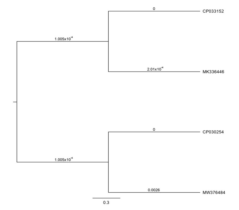
Figure 4. Phylogenetic tree analysis relied on the ITS1 gene-specific region of Malasseziarestrictafrom Iraq (MW376484); their host is a wild cat. Sequencing revealed close matching of the phylogenetic tree to an isolate from Korea (CP030254). The compression was performed using NCBI – the based nucleotides website.
CONCLUSIONS
The current study spotlighted the species of Malasseziarestricta that infected domestic and wild cats by molecular analysis for the first time in Iraq, which was inserted in GenBank: MW376484.1 from wild cat Felischausfurax. This is a significant result as it proves that the fungus Malasseziarestricta is a common infection between humans and animals (zoonotic infection) and that cats can play this role.
Ethical approval
The trial was registered in "The Iraq Natural History Research Center and Museum INHM" (Email: [email protected] ). The research proposal was approved by the Scientific Affairs Department of Baghdad University (SH.A.923/17/2/2021).
Conflicts of interest: The authors declare no conflict of interest related to the work in a manuscript.
REFERENCES
1-GBIF, Global Biodiversity Information Facility, 2021. https://www.gbif.org/species/2518093
2-Guého, E., Midgley, G. and Guillot, J. ,"The genus Malassezia with description of four new species", Antonie van Leeuwenhoek , 1996 (69): 337–355.
3-Thayikkannu, A. B. ,Kindo, A. J., andVeeraraghavan,M., " Malassezia-Can it be ignored", Indian JDermatol, 2015;( 60): 332-339
4-Levin, N.A., and Delano, S., " Evaluation and treatment of Malassezia
related skin disorders", Cosmet. Dermatol. , 2011; (24):137‑145
5-Ran, Y., "Observation of Fungi, Bacteria, and Parasites in Clinical Skin Samples Using Scanning Electron Microscopy". In Janecek, Milos; Kral, Robert (eds.). Modern Electron Microscopy in Physical and Life Sciences.InTech. 2016.
6-Bond,R., Daniel, O., Morris, J. G., Emmanuel, J., Bensignors, D. R., Kenneth, V. M., Rui, K., and Peter B. H.,"Biology, diagnosis and treatment of Malassezia dermatitis in dogs and cats Clinical Consensus Guidelines of the World Association for Veterinary Dermatology, Veterinary Dermatology, 2020; 31 (27 –e4):1-49.
7-Crespo, M. J.,Abarca, M. L., and Caban˜es, F. J., "Isolation of Malassezia furfur from a Cat", Journal of Clinical Microbiology, vol. 37, no.5, pp. 1573–1574, 1999.
8-Morris, D.O., O'Shea, K., Shofer, F.S., Rankin, S., "Malasseziapachydermatis carriage in dog owners" , Emerg. Infect. Dis. [serial on the Internet].,2005:11( 1): 83-88.
9-Bożena, D. K., Małgorzata, J. B., and Iwona, D., "Occurrence of various pathogenic and opportunistic fungi in skin diseases of domestic animals: a retrospective study", BMC Veterinary Research, 2020;(16): 248 .
10-Khosravi, A., Eidi, S., Katiraee, F., Ziglari, T., Bayat, M., Nissiani, M., "Identification of different Malassezia species isolated from patients with Malassezia infections" , World Journal of Zoology, 2009; 4(2): 85-89.
11-Sugita, T., Mami, T., Misato, A., Ryoji, T., and Akemi, N. , "Genotype Analysis of Malasseziarestrictaas the Major Cutaneous Flora in Patients with Atopic Dermatitis and Healthy Subjects" Microbiol. Immunol., 2004;48( 10): 755–75.
12-Annabelle, M., Haydar, F., Alice, C., and John, V. W. "Prolonged Fever and Splenic Lesions Caused by Malasseziarestrictain an Immunocompromised Patient", Author manuscript, Pediatr Transplant, 2014; 18( 8): 283-286.
13-Al-Jabry, A. T., and Alsudani, A. A., "Survey of Malasseziaspp. that causing PityriasisVersicolor in Al-Diwaniyah city, Iraq", European Journal of Molecular & Clinical Medicine, 2020;7(2): 4416- 4428 .
14-Awad, A.K., Al-Ezzy, A. I., and Jameel, GH., " Phenotypic Identification and Molecular Characterization of Malassezia Spp. Isolated from PityriasisVersicolor Patients with Special Emphasis to Risk Factors in Diyala Province, Iraq", Open AccessMaced. J.Med. Sci., 2019 ; 7(5):707-714.
Received: December 23, 2022 / Accepted: January 30, 2023 / Published:15 February 2023
Citation: Muslim Hadi A, Saber Khalif H. Molecular analysis of Fungi: Malasseziarestricta from Felidae. Revis Bionatura 2023;8 (1)46. http://dx.doi.org/10.21931/RB/2023.08.01.46