2020.05.03.20
Files > Volume 5 > Vol 5 No 3 2020
NEWS AND VIEWS /NOTICIAS Y OPINIONES
Use of natural diatoms for drug delivery

Castro Dario1, Cuasquer Joselyn2, Chavez
Available from: http://dx.doi.org/10.21931/RB/2020.05.03.20
ABSTRACT
Diatoms are microalgae organisms that have a cover of silica, with a fascinating ordered porous structure that varies in size, giving them some different characteristics. Because of their different size, shape, and structure, it has incredible properties, letting them be capable of been functionalized with other particles. Therefore, due to the ordered pore structure, the high surface area, biocompatibility, availability, and low processing cost, they present a growing potential for drug delivery when talking about silica materials, natural and synthetic, not to mention that is less expensive and a green alternative.
Keywords: Diatoms; Biosilica; Surface functionalization
INTRODUCTION
Diatoms are unicellular microalgae that are the most outstanding natural source of porous silica; these species can be found in every aqueous habitat; there exist probably around 100000 species of them. Nowadays, diatoms are marveling nanotechnologists who are hoping these amazing pore structures can show them how to improve minute structures to do better tasks.
The first time that the idea of build new biological materials by the structure of the diatom came in 1999 where scientists realize the potential that present diatoms due to the nano-scale design and the genetic control, essential features for the human engineering1.
During their accelerated growth cycle, diatoms absorb substantial amounts of trace elements and nutrients from the surface water layer, especially silicon to form their shells, and zinc, which plays a vital physiological role in their development2.
Morphology of Diatoms
The diatoms show a very diverse and successful lineage of photosynthetic Stramenopiles (Chromalveolates) with cell walls formed by amorphous silica and consisting of two parts, termed frustules, as their most striking feature. Their high abundance, coupled with the resilience of diatom frustules to disintegration in healthy waters, has resulted in massive sedimentary aggregation and a significant fossil record. The diatoms, as mentioned before, are unicellular beings, but some form colonies. The diatom plastids are derived from red algal secondary symbiosis and are golden brown due to the high concentration of the carotenoid fucoxanthin3. Diatoms were abounding and believed to be the most important group of eukaryotic phytoplankton, in charge of almost 40% of principal marine productivity4.
Reproduction of diatoms
Diatoms reproduce by two different modes, sexual and asexual. The ability to reproduce sexually is associated with the cell size of the diatom. It is the most pervasive strategy for re-establishing cell size5. A male gametangial cell initially experiments a progression of separated divisions to shape a number set of sperm mother cells, at that point, experience meiosis to frame flogged sperm. Oogonial female cells are capable of producing just one or even a maximum of two eggs, and then the fertilization forms a zygote6; on the other hand, Diatoms have a unique mode of asexual reproduction. The vegetative cell division involves a successive reduction in the mean cell. New sibling valves are usually formed in close juxtaposition to each other. There are many consequences because of this, and as an example is a fact that diatoms must be able to escape from the inevitable size diminution if they are to survive, it means that with the time they must use the sexual reproduction to recover standard size6.
Types of diatoms
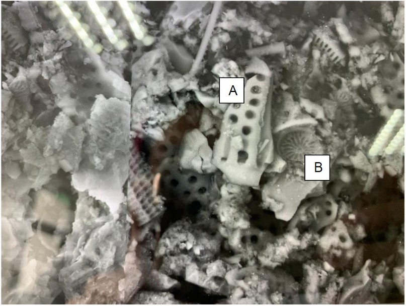
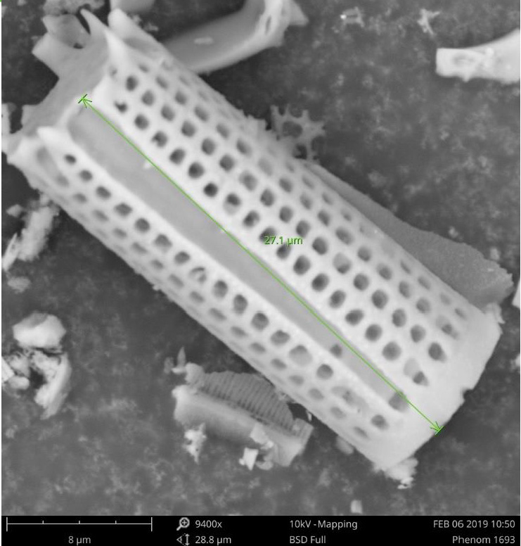
Diatoms have unique features, and good structural architecture made-up of silica provides various opportunities for the design of new materials7. More than 10 000 species of diatoms are known, and they are characterized based on the shape and structure of their cell walls, the diatom's cell wall structure can be a hexagonal, rod and circular shape8. There are two major types of diatoms, centric (Figure 1) and pennate, which are easily differentiated by frustule symmetry. Pennate diatoms tend to be elongated and centric diatoms are radially symmetrical. The centric type is of primary interest as having nano-engineering potential. The cylindrical type (Figure 2), its uniform pore structure, well-aligned pores, and wide ranges of heights and diameters9.

Figure 1. Microscopic picture showing both types of diatoms (A) cylindrical and (B) centric.

Figure 2. Cylindrical diatom looked by Scanning Electron Microscope (SEM)
Chemical composition of diatoms
Two types of silicon are found: condensed silica (SiO2) and weakly polymerized silicate species (inorganic silicate, or silicate ester). The chemical composition of the diatom surface contains many lipids as peptides, polysaccharides, hydrocarbon-like compounds, also sort of SiO2, and Mg(OH)2. XPS analysis reveals a high concentration of lipids in the form of carboxylic esters; additionally, the organic part of the walls contains protonated nitrogen and phosphate. They are attributed not only to phospholipids but also, and possibly mainly, to phosphopeptides already described in diatom frustule, like silaffins or silacidins, also in walls Sulfate groups are also detected and are attributed to monoesters of polysaccharides10.
Applications of diatoms
Diatoms have a considerable quantity of applications in different areas. Nanotechnology is one of the fields where these marvelous microalgae have considerable applications worth to mention such as Bio-mineralization, which is a natural method where biosystems are used to extract minerals that derive from organic compounds and can be assisted by micro-organisms including diatoms, bacteria, and fungus11. Diatoms are used more often than the others because of their large number of individuals and also by the capability of produce biosilica, which is an essential mineral for their sructure11, 12.
The unique structure of the frustules in diatoms allows us to use them in water filters, building materials, chromatography supports, and also cadmium production because this metal is a waste in the development of diatoms12. The improvement in the nanotechnology field encourage the production of smaller and efficient pieces to assemble electronic, optical, chemical, or biomedical devices13.
Industries nowadays produce much more pollutants that are released in freshwater at the environment that create a considerable adverse alteration in the physical, chemical, and biological properties of the water11. For this reason, all the pollutants in the environment must be managed and controlled. Biological methods for pollutants involve techniques to reduce their toxicity in the environment by transforming and mineralization of these pollutants. Diatoms are used in biodegradation of pollutants due to their quick response of disturbances in the environment and also their structure. There is evidence that certain diatoms species are capable of working as biological indicators for pollutants in rivers11.
Diatoms had formed a 2D array, thus bringing up their use in biodevices development. Freshwater diatoms have been used to make biosensors for water quality assessment using alternating current dielectrophoresis to chain live diatom cells to create a 2D array 14. Nanomedicine and medical applications employ nanomaterials, nano electrical biosensors, and molecular nanotechnology with drug delivery vehicles, diagnostic devices, and physical therapy applications being all-important in the general panorama. The big problem nanomedicine has to deal with is toxicity, biodegradability, and environmental impact. Using diatoms or their derived frustules instead provides intricate homogeneity while also surpassing the shortcomings as they are non-toxic, biodegradable, and readily available in the environment. Some of these applications are biosensors, immunodiagnostics, optical biosensors, and drug delivery14.
Mesoporous silica nanoparticles can be produced with several variations of size, morphology and tunable properties, the last one associated with specific applications, as mentioned before in the introduction, the synthesis of these materials includes complex chemical processes as well as toxic and expensive reagents. The characteristics that a diatoms' silica shells possess can excel the ones shown in the existing synthetic silica nanoparticle delivery systems, some of them: the frustules can be easily functionalized, protected, and engineered for controlled drug loading and successive delivery through the nanopores. In 2016, Ragni et al. mentioned the work of other authors that demonstrated the potential of diatom silica as a rising biomaterial for the delivery of a certain desired drug to a specific site.15
Drug delivery system
Drug delivery is one of the technological advances whose main objective is the administration or release of medications in the individual. The use of medications is a common practice in human history, but how medications are administered has varied and improved over time, being much more efficient16. Thanks to the use of biotechnology, we have been able to generate more specific and compound medicines, making their administration more intricate. The most modern systems include the use of microspheres and polymer microcapsules, nanoparticles, and natural polymers17.
In order to consider a specific type of drug delivery, it is necessary to consider the clinical needs and the factors that characterize them, such as the physicochemical properties of the drug, the disease to be treated, in which part of the body has its interaction if the treatment will be for chronic or acute diseases, among others17. According to the National Institute of Biomedical Imaging and Bioengineering of the USA, the most recent drug delivery systems research can be described in 4 different categories such as administration routes, administration vehicles, cargo, and mark strategies16.
Controlled delivery systems
Controlled systems are used to maintain adequate levels of drugs in the body, which is necessary for the administration of certain medications making their use more efficient. The main objective for which this system was conceived is to maintain constant levels of medications in the patients' blood for a while, thus avoiding a decrease in the concentration of medication between administrations, ensuring that the dose does not reach toxic levels for the patient18. Chemical-controlled systems of the bioerosion or biodegradation system, which consists of the degradation of the polymers where the medicine is distributed in water-soluble molecules, can also use the system Pendant chain of a biodegradable polymer which, upon contact with the water destroys these bonds by releasing the medication in the body19.
Diatoms for drug delivery system
One of the main objectives in drug delivery is to develop new and efficient carries for drugs with less toxicity for the patient and reduce the risk of manage high amounts of drugs incorporations20. Through the years, several experiments had been made facing the struggles for drug delivery, and as a result, bio-templating was developed and one of them came from diatoms21. The diatom nanostructure has adequate characteristics such as absorption capacity and packing for drug delivery23. Using diatoms for drug delivery allows us to understand the effect of surface functionalization on controlling diffusion rates and drug delivery due to the drug packing and release in bare diatoms and modified diatoms1.
Properties of diatoms in a drug delivery system
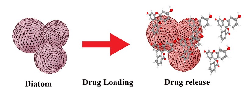
Diatom shells possess a combination of chemical, mechanical, and structural characteristics that surpass the obstacles associated with the delivery of therapeutic agents and offer several advantages over existing synthetic microparticle delivery systems. They have a pillbox structure with a hollow and sizeable inner space with micro- and nano-scale porosity, high surface area, excellent biocompatibility, amorphous silica, high permeability, low density, non-toxicity and the ability to mimic the nature of constituents for natural bone and medical implant, this make diatom silica a promising biomaterial for drug-delivery applications23 (Figure 3). Diatoms with distinct 3-d of silica cell walls and highly-ordered pore structures, offering a great potential to replace synthetic mesoporous materials also as suitable carrier system used porous diatomite nano-carriers for delivering small interfering ribonucleic acid (siRNA) inside the human epidermoid cancer cells 24.

Figure 3. Diatom as a drug delivery system. The drug is loaded inside the silica frustule of the diatom. Once the encapsulation is exposed to the right environmental conditions, its content will get discharged into the media.
Diatom's frustule
The diatoms have an outer wall composed of silica called frustule which consists of two leaflets, a small one called hypotheca and a larger one called an epitheca, each of which is formed from a leaflet that forms the outer surface and a girdle that is a circular band of silica on the edges of the valve3. These leaflets are located from different sheets, which gives them a variation in porosity that can range from microns to nanometers, and their distribution depends on the species. The use of frustules diatoms has varied over time and can be found in these physical properties that allow its application in fields such as nanotechnology and biotechnology, where we can highlight its use for the production of bio-inspired solar cells due to its capacity of collecting light with high efficiencies, such as nanostructured substrates in plasmonics, optical sensors, and biosensors, nanoparticles as vectors in drug delivery. It is possible to generate variations in the frustule diatoms in terms of their porosity, morphology, and geometry to be able to generate new, more efficient techniques; this is possible through the modification of specific genes that are involved in the formation of the frustule diatom25.
Dolatabadi and de la Guardia exposed some studies about the functionalization of diatom biosilica frustules with antibodies and enzymes26. Trying to take out a new characteristic of diatoms, it is known that diatom-silica microcapsules were functionalized with dopamine-modified iron-oxide NPs. Then a new property to use diatoms was born, the magnetic diatoms, which by the way, have an enormous potential to be used as magnetically-guided drug-delivery micro-carriers.
Separation and purification of diatom biosilica
It is essential for the use of diatoms to purify the samples of any impurity, such as clay and organic substances. These substances are present in the diatoms because the environment and the conditions that were exposed the diatoms for their development27.
For the purification process, we can take diatoms directly from freshwater and put them for two weeks in a growth medium rich in silicate for their treatment with piranha solution to remove organic waste28, pulverize the samples using Milli Q water followed by sonication, filtration and sedimentation processes24, 29.
Diatoms as drug carriers
The use of silica-based particles has been increasing over the years creating a wide range of porosities and scales for drug delivery; these synthetic particles have the perfect characteristics such as a wide surface area, thermal stability, biocompatibility, among others. However, their high cost, the unnecessary production time and the use of toxic materials have made it necessary to look for other natural sources of these silica structures for the drug delivery, with diatoms being the best positioned with their exceptional characteristics30.
In-vitro tests and experiments have been made in which the diatoms take an essential role as the carriers of certain drugs, such as the indomethacin with which it was possible to demonstrate loading capacity (22 wt%) and prolonged drug release over two weeks giving promising results in the use of diatoms30. Other drugs tested in diatoms were mesalamine and prednisone both are common drugs to the treatment of gastrointestinal disorders, the drug delivery property of diatoms with these two drugs where tested in monolayers of colon cancer cells and demonstrate low toxicity at a concentration of 1000 µg/ml and also a stable release of drugs in a period; moreover, the study prove that the diatoms enhance the permeability of the drug by the permeation of the monolayer cell culture31.
CONCLUSION
The use of diatoms for drug delivery presents a new option in the field. However, studies are still lacking31; the results that have been obtained to date are very encouraging, opening new research areas and generating a viable solution to the use of silica structures synthetic, which have high production costs and use toxic materials in their production. Diatoms are one of the many natural resources that we have within our reach to be able to develop better and more efficient methods of drug delivery since these present exceptional characteristics to package and release drugs inside; we have before us a great opportunity which it needs to be further researched and developed.
REFERENCES
- Chao, Joshua T, Manus JP Biggs, and Abhay S Pandit. 2014. "Diatoms: A Biotemplating Approach to Fabricating Drug Delivery Reservoirs." Expert Opinion on Drug Delivery 11 (11)
- Vance, Derek, Susan H. Little, Gregory F. De Souza, Samar Khatiwala, Maeve C. Lohan, and Rob Middag. 2017. "Silicon and Zinc Biogeochemical Cycles Coupled through the Southern Ocean." Nature Geoscience 10 (3): 202–6.
- Brinkmann N., Friedl T., Mohr K.I. (2011) Diatoms. In: Reitner J., Thiel V. (eds) Encyclopedia of Geobiology. Encyclopedia of Earth Sciences Series. Springer, Dordrecht.
- Falkowski, Paul G, Richard T Barber, Victor Smetacek, Paul G Falkowski, Richard T Barber, and Victor Smetacek. 1998. "Linked References Are Available on JSTOR for This Article : Biogeochemical Controls and Feedbacks on Ocean Primary Production." Science 281 (5374): 200–206.
- Gandhi, H.P. 1966. Freshwater diatom flora of Jog Falls, Mysore State. Nova Hedwigia, 11: 89-197.
- Gandhi, H. P., A B. Vora and D. J. Mohan, 1983. Fossil diatoms from Baltal, Karewa beds of Kashmir. Current Trends in Geology, Vol. VI, (Climate and Geology of Kashmir) 6:61-68.
- A. Jantschke, A.K. Herrmann, V. Lesnyak, A. Eychmuller, E. Brunner, Decoration of diatom biosilica with noble metal and semiconductor nanoparticles (<10 nm): assembly, characterization, and applications, Chem. - Asian J. 7 (2012) 85-90.
- K.M. Wee, T.N. Rogers, B.S. Altan, S.A. Hackney, C. Hamm, Engineering and Medical Applications of Diatoms, J. Nanosci. Nanotechnol. 5 (2005) 88-91.
- Y.S.S. Sofia Sotiropoulou, Sonny S. Mark, and Carl A. Batt, Biotemplated Nanostructured Materials, Chem. Mater. 20 (2008) 821-834
- Tesson, B., M. J. Genet, V. Fernandez, S. Degand, P. G. Rouxhet, and V. Martin-Jézéquel. 2009. “Erratum: Surface Chemical Composition of Diatoms (ChemBioChem (2009) Vol. 10 (2011-2024)).” ChemBioChem 10 (12): 1915.
- Jamali, Ali Akbar, Fariba Akbari, Mohamad Moradi Ghorakhlu, Miguel de la Guardia, and Ahmad Yari Khosroushahi. 2012. "Applications of Diatoms as Potential Microalgae in Nanobiotechnology." BioImpacts 2 (2): 83–89.
- Jiao, Jun, Timothy Gutu, Debra K. Gale, Clayton Jeffryes, Wei Wang, Chih Hung Chang, and Gregory L. Rorrer. 2009. "Electron Microscopy and Optical Characterization of Cadmium Sulphide Nanocrystals Deposited on the Patterned Surface of Diatom Biosilica." Journal of Nanomaterials 2009.
- Gordon, Richard, Frithjof A.S. Sterrenburg, and Kenneth H. Sandhage. 2005. "A Special Issue on Diatom Nanotechnology." Journal of Nanoscience and Nanotechnology 5 (1): 1–4.
- Mishra, Meerambika, Ananta P. Arukha, Tufail Bashir, Dhananjay Yadav, and G. B.K.S. Prasad. 2017. "All New Faces of Diatoms: Potential Source of Nanomaterials and Beyond." Frontiers in Microbiology 8 (JUL): 1–8.
- Ragni, Roberta, Stefania Cicco, Danilo Vona, Gabriella Leone, and Gianluca M. Farinola. 2017. “Biosilica from Diatoms Microalgae: Smart Materials from Bio-Medicine to Photonics.” Journal of Materials Research 32 (2): 279–91.
- Nibib.nih.gov. (2019). Drug Delivery Systems | National Institute of Biomedical Imaging and Bioengineering. [online] Available at: https://www.nibib.nih.gov/node/42846
- S.S. Davis (2000) Drug delivery systems, Interdisciplinary Science Reviews, 25:3, 175-183
- Bhowmik, D. et al. (2012). Controlled Release Drug Delivery Systems. The Pharma Innovation. Available: http://www.thepharmajournal.com/vol1Issue10/Issue_dec_2012/3.1.pdf
- Dr. Liji Thomas, M. (2019 Chemically Controlled Drug Delivery Systems. from https://www.news-medical.net/health/Chemically-Controlled-Drug-Delivery-Systems.aspx
- Venkatesan, Jayachandran, Baboucarr Lowe, and Se-kwon Kim. 2015. "Application of Diatom Biosilica in Drug Delivery," 245–54.
- Maher, Shaheer, Tushar Kumeria, Moom Sin Aw, and Dusan Losic. 2018. "Diatom Silica for Biomedical Applications : Recent Progress and Advances" 1800552: 1–19.
- Farinola, Gianluca M, Stefania R Cicco, Dipartimento Chimica, Bari Aldo, and Moro Via. 2017. “Diatoms Biosilica as Efficient Drug-Delivery System,” 3825–30.
- Aw, Moom Sinn, Spomenka Simovic, and Jonas Addai-mensah. n.d. "Silica Microcapsules from Diatoms as New Carrier for Delivery of Therapeutics P Reliminary C Ommunication," 1159–73
- .Bariana, Manpreet, Moom Sinn Aw, Mahaveer Kurkuri, and Dusan Losic. 2013. "Tuning Drug Loading and Release Properties of Diatom Silica Microparticles by Surface Modifications." International Journal of Pharmaceutics 443 (1–2): 230–41.
- De Tommasi, E., et al., Diatom Frustule Morphogenesis and Function: a Multidisciplinary Survey, Mar. Genomics (2017), http://dx.doi.org/10.1016/j.margen.2017.07.001
- Dolatabadi, Jafar Ezzati Nazhad, and Miguel de la Guardia. 2011. "Applications of Diatoms and Silica Nanotechnology in Biosensing, Drug and Gene Delivery, and Formation of Complex Metal Nanostructures." TrAC Trends in Analytical Chemistry 30 (9): 1538–48. https://doi.org/10.1016/j.trac.2011.04.015.
- M.S. Aw, S. Simovic, Y. Yu, J. Addai-Mensah, D. Losic, Porous silica microshells from diatoms as biocarrier for drug delivery applications, Powder Technol. 223 (2012) 52-58
- N.L. Rosi, C.S. Thaxton, C.A. Mirkin, Control of nanoparticle assembly by using DNA-modified diatom templates, Angew. Chem. 43 (2004) 5500-5503.
- I. Rea, N.M. Martucci, L. De Stefano, I. Ruggiero, M. Terracciano, P. Dardano, N. Migliaccio, P. Arcari, R. Tate, I. Rendina, A. Lamberti, Diatomite biosilica nanocarriers for siRNA transport inside cancer cells, Biochim. Biophys. Acta, 1840 (2014) 3393-3403.
- Aw, Moom Sinn, Spomenka Simovic, Jonas Addai-Mensah, and Dusan Losic. 2011. "Silica Microcapsules from Diatoms as New Carrier for Delivery of Therapeutics." Nanomedicine 6 (7): 1159–73.
- Zhang, Hongbo, Mohammad Ali Shahbazi, Ermei M. Mäkilä, Tiago H. da Silva, Rui L. Reis, Jarno J. Salonen, Jouni T. Hirvonen, and Hélder A. Santos. 2013. "Diatom Silica Microparticles for Sustained Release and Permeation Enhancement Following Oral Delivery of Prednisone and Mesalamine." Biomaterials 34 (36): 9210–19. https://doi.org/10.1016/j.biomaterials.2013.08.035.
Received: 20 June 2020
Accepted: 20 July 2020
Castro Dario1, Cuasquer Joselyn2, Chavez
Castro Dario1 https://orcid.org/0000-0002-3194-9601,
1. School of Biological Science and Engineering Yachay Tech
Cuasquer Joselyn2 https://orcid.org/0000-0002-3510-9976 ,
2. School of Biological Science and Engineering Yachay Tech
Chavez
3. School of Biological Science and Engineering Yachay Tech
Corresponding author: [email protected]