2023.08.01.9
Files > Volume 8 > Vol 8 No 1 2023
Protocol for histological analysis of the interaction Vanilla planifolia - Fusarium oxysporum f. sp. vanillae

Quirino Villarreal Alejandro1 , Ramírez Vázquez Mónica2, Velázquez López Olinda Elizabeth3
 , Rivera-Fernández Andrés1, Luna-Rodríguez Mauricio1*
1Laboratorio de Genética e Interacciones Planta Microorganismos, Facultad de Ciencias Agrícolas, Universidad Veracruzana. Circuito Gonzalo Aguirre Beltrán s/n, Zona Universitaria, Xalapa, Veracruz, CP 91000, México. e-maiL: [email protected].
2 Facultad de Ciencias, Universidad Nacional Autónoma de México (UNAM), Circuito Exterior, Cd. Universitaria, Copilco, Coyoacán, Ciudad de México 04510, México. [email protected].
3Instituto de Ecología A.C. El Haya, Xalapa, Veracruz, CP 91073, México. [email protected].
* Correspondence: [email protected].
Available from: http://dx.doi.org/10.21931/RB/2023.08.01.9
ABSTRACT
Fusarium oxysporum f. sp. vanillae (Fov) is the main phytosanitary problem of Vanilla planifolia, invading the root and causing the rotting of up to 80% of the plants. The histological process is used to study the morphological characteristics of the tissues of interest and published a protocol to check Fov-V. planifolia interaction that was not reproducible under our conditions, so the study aimed to adapt a procedure that allows differential analysis of the mechanism of Fov invasion in V. planifolia by confocal microscopy. The bioassays consisted of analyzing Fov-infected roots. The experimental design consisted of three replicates evaluated at 12, 24, 36, 48, 60 and 72 hours post-inoculation. Roots without inoculum were used as controls. The samples were fixed in paraformaldehyde and 4% sucrose (PBS). Subsequently, the tissues were dehydrated by a combination of alcohols at different concentrations. The cuts were made with a microtome. The sections were observed under a Leica confocal microscope with a 63X objective with ap. No. 1.4, plus immersion oil; excitation lines: 405, 488, 532. The FAA + kerosene method was the procedure that allowed complete tissue sections.
Keywords: Fusariosis, Vanilla, interaction.
INTRODUCTION
Vanilla planifolia Jacks. is one of the few orchids in widespread commercial use. Its pods are essential for the manufacture of vanilla flavoring. Because of its clonal propagation, V. planifolia has been reported to have low genetic diversity, which makes it susceptible to diseases. In particular, root and stem rot caused by Fusarium oxysporum f. sp. vanillae, is the result of the penetration of hyphae into the root tissues, which in severe cases causes tissue death and, consequently, the death of the plant1. Fusarium has caused the loss of entire crops and represents a critical point for current production systems.
Studies of the V. planifolia - F. oxysporum interaction are still limited. Most of them focus on the evaluation of physiological aspects of the stem, leaves and fruits, giving less importance to the interaction with the roots 1, 2, 3, 4. Vascular symptoms, such as colonization and dissemination of the causal agent in root tissues, have been ambiguously described 5.
Improving methods that help to understand the basis of this interaction is of utmost relevance for developing disease control strategies. The histological process is a series of methods and techniques used to study the morphological and molecular characteristics of the tissues of interest, depending on what is to be observed and the type of microscopy to be used. Since the technical requirements of the proposed methods for the study of this interaction were not available, it was proposed to establish an alternative histological procedure that allows differential analysis of the mechanism of invasion of F. oxysporum f. sp. vanillae in the cellular structures of the root tissue of V. planifolia using confocal microscopy.
MATERIALS AND METHODS
Bioassays. The V. planifolia material was collected in an orchard established in the town of Aparicio in the municipality of Vega de Alatorre, Veracruz. According to Solano-de la Cruz et al. (2019)5, root generation was induced in vanilla cuttings. To promote the invasion of Fusarium oxysporum f. sp. vanillae (Fov) in V. planifolia tissues, aqueous suspensions of spores of strain HG3C1 inoculated in the zone of absorbent root hairs were used. Previously, the fungus was activated on potato dextrose agar (PDA) medium for ten days. After inoculation, the roots were incubated at 28 2 ºC in a humid chamber for 72 hours, with observations every 12 hours, until the appearance of the initial symptoms of the disease was detected.
Prefixation of the samples. After detecting the initial symptoms of the disease, the roots were cut and prefixed in paraformaldehyde and 4% sucrose solution (PBS) in a 1:10 ratio (weight/volume) 6. Subsequently, three washes were performed by immersion in sterile distilled water at one-hour intervals, changing the water at each immersion. To ensure PBS removal, the samples were observed under a light microscope. Once the PBS was removed, the samples were immersed in FAA fixative solution (90 mL of 70% ethyl alcohol, 5 mL of formaldehyde and 5 mL of acetic acid) for 24 h. The samples were then immersed in the FAA fixative solution (90 mL of 70% ethyl alcohol, 5 mL of formaldehyde and 5 mL of acetic acid) for 24 h. The fixative solution was then removed by immersing the samples in sterile distilled water, with changes every 30 min7, 8. The examples were dehydrated by immersion in alcoholic solutions (ABT) (Table 1). In the first instance, solutions ABT1 to ABT5 were used, with immersion times of 30 min. Subsequently, they were immersed in the ABT6 solution for at least 2 hours if the sample was used immediately for mounting and cutting; otherwise, it was left engaged in ABT6 until it was used for climbing, not exceeding 24 hours.
Mounting of the samples. The following methods were tested for the mounting stage: a) Mounting in kerosene. The samples were dried in a heating oven at 60 °C. Histological kerosene was heated to the liquid phase and poured into 1.5 cm3 cubic cardboard molds. While filling the molds, the root was placed in a horizontal orientation and kerosene was added until the tissue was covered entirely. It was left to solidify at room temperature for one hour; b) Mounting in freezing with paraplast. The roots were placed horizontally in a plastic mold, and liquid paraplast was added. The mold was placed in a cryostat for freezing; c) Mounting with paraplast. The sample was placed horizontally in a plastic mold, and liquid paraplast was added. It was left to solidify at room temperature for one hour; d) Mounting with polyethylene glycol. Cubic cardboard molds were filled with liquid polyethylene glycol. Root samples were placed horizontally and allowed to stand at room temperature for one hour; e) Mounting with FAA + kerosene. The samples were placed horizontally in plastic molds and dried in an oven at 60 °C until complete evaporation of the FAA; then, liquid kerosene was added and allowed to stand at room temperature for 30 minutes until solidification.
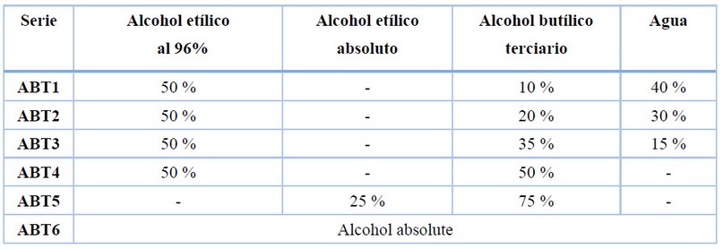
Table 1. Concentration of alcohols used for dehydration of V. planifolia Jacks. root samples.
Cutting. Two techniques, microtome and cryostat, were used to obtain root tissue sections. For this, a Leica slide microtome (Mod. SM2010 R) with thickness settings at 10, 20, 40 and 60 µm 1 was used; for cryostat cuts, a Leica Biosystems (Mod. CM 1860 UV) with thickness settings at 10, 20, 40 and 60 µm, and temperature settings at -13, -15 and -21 °C was used.
Sample recovery and staining. Tissue sections obtained with cryostat were placed directly on the slides for staining. For the separation of kerosene, PEG and paraplast from tissue sections obtained with a microtome, these were immersed in an aqueous solution of 1 g/L porcine skin gelatin type A at 60 °C. A Barnant flotation bath (Mod. MH8517) was used to maintain a constant temperature. After separation, the sections were placed on slides. Tissue integrity was checked under a compound microscope at 40X.
In all cases, staining was carried out with fluoride tracing white (5 min) and then propidium iodide (5 min) 9. After adding the dyes, the slides were placed at 60 °C, two to three drops of ethanolic solutions from 96, 70, 50 and 30 % were gradually administered, and finally, sterilized distilled water for tissue rehydration 1.
Microscopic observation. A Leica confocal microscope (SP8 STED) with 63X objective. ap. no. 1.4 + immersion oil and 405, 488, and 532 nm excitation lines were used.
RESULTS
Cryostat slices. Samples 40 and 60 µm thick from all mounting methods (kerosene, paraplast, paraplast freezing, PEG and FAA+paraffin) were inadequate to differentiate Vanilla root and fungal structures (Figure 1 A, a). Samples 10 and 20 µm thick did not show tissue integrity (Figure 1 B, b and C, c).
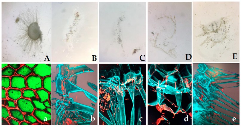
Figure 1. Appearance of images from histological sections of F. oxysporum f. sp. vanillae-infected V. planifolia roots processed by the different mounting and cutting techniques tested. A, thick aspect slice; B and C, fragmented slices obtained with cryostat; D and E, fragmented slices obtained with a microtome. a, b, c, d and e, confocal microscopy images of the pieces referred to in A, B, C, D and F, obtained with 63X objective. ap. no. 1.4, immersion oil and excitation lines 405, 488 and 532. Photos: Quirino, V. A., 2021.
Microtome slices. Only the 10 and 20 µm thick samples from the FAA + kerosene mounting method were intact and of good quality for microscopic observation (Figure 2a). The internal tissues of vanilla root were differentially recognized by the blue coloration, produced by the reaction of the white reagent fluorine chalcogenate with the plant cell walls, and the red coloration produced by the interaction of propidium iodide with the fungal hyphae (Figure 2b).
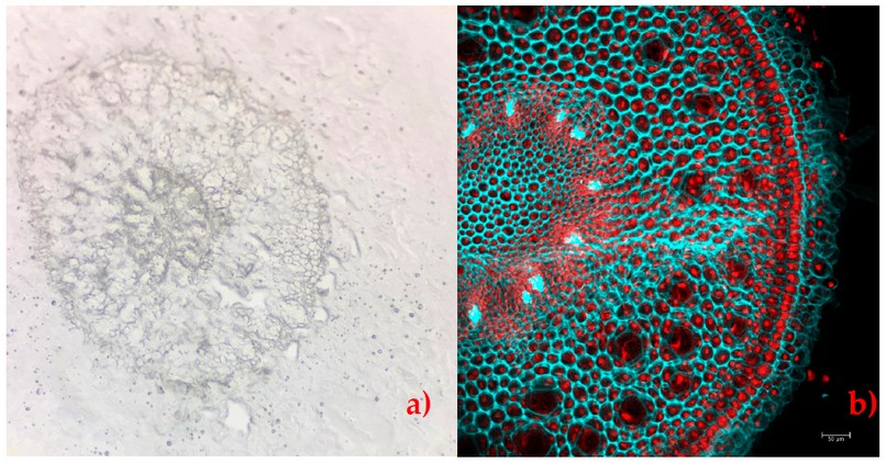
Figure 2. Appearance of V. planifolia root sections with FAA+ paraffin mounting made with microtome at 10 µm thickness. a) verification of tissue integrity with compound microscopy; b) root stained and visualized in confocal microscopy with 63X objective. ap. no. 1.4, immersion oil and excitation lines 405, 488 and 532. Plant cells are shown in blue, and F. oxysporum f. sp. vanillae colonization is in red. Photos: Quirino V.A., 2021
DISCUSSION
The use of histological techniques makes it possible to observe structures, in a general or detailed manner, of the different components of a sample; therefore, they have been useful for studying the behavior of pathogens at the tissue and cellular level, which has made it possible to make timely diagnoses10, 11, 12. A complementary tool to histological techniques is confocal scanning optical microscopy, where the operation of such a microscope is similar to that of an epifluorescence microscope since both are based on the phenomenon of fluorescence; however, the latter provides images of poorer quality. This is because epifluorescence microscopes show information from all planes of the sample, focused or not, because they lack the confocal detection diaphragm and use a mercury lamp as an illumination source, which has a very irregular power, with different intensity peaks 13.
Although, for the study of the pathosystem V. planifolia - F. oxysporum, they reported an investigation where they used epifluorescence microscopy and multiphoton microscopy with different techniques of mounting and staining of the samples; however, some images are far from being quick decipherable1. In addition, not all laboratories have the technical and instrumental elements necessary to reproduce the method, hence the importance of adapting alternative ways that do not limit the research.
In the case in question, the confocal microscopy and the histological techniques used allowed for obtaining clear images, which facilitated the differentiation of the cellular structures corresponding to each organism; by staining the cells of the vanilla root in blue color due to the affinity of the white fluoride tracing reagent with the lignin and the fungus cells in red color due to the affinity of the propidium iodide reagent with the chitin 9. Additionally, it was observed that only mounting with FAA + kerosene maintained the integrity of vanilla root tissue upon sectioning. FAA is a widely used fixative for soft plant tissues, such as vanilla root, whose advantages include rapid and complete penetration of formaldehyde into the tissue, prolonged storage possibilities, and a balance of alcohol and acid shrinkage/expansion. In addition, formaldehyde is a non-coagulation fixative that chemically binds cellular components to preserve the structure 14. For its part, polyethylene glycol acts as a barrier by blocking the entry of other substances into the tissue 15, which could have caused the lack of rigidity of the vanilla root to remain intact at the time of cutting.
CONCLUSIONS
A histological method based on FAA + kerosene for sample mounting and confocal microscopy was adapted to differentiate the cellular structures of Vanilla planifolia root and those of Fusarium oxysporum f. sp. vanillae during their interaction in the development of the disease known as Vanilla root and stem rot.
Author Contributions: Generation of the idea and resources: Mauricio Luna Rodríguez, Alejandro Quirino Villarreal and Mónica Ramírez Vázquez. Methodology: Alejandro Quirino Villarreal, Mónica Ramírez Vázquez, Olinda Elizabeth Velázquez López and Mauricio Luna Rodríguez. Concepts: Mauricio Luna Rodríguez, Andrés Rivera Fernández and Mónica Ramírez Vázquez. Preparation of the original draft: All authors.
Funding: This research was funded by CONACYT-FORDECYT 2018 (Project 29748, "Adaptation and mitigation strategies to climate change, necessary for the rescue of vanilla cultivation in Mexico").
Acknowledgments: To Dr. Guillermo Ángeles Álvarez, Instituto de Ecología, A.C. To the Institutional Fund for Regional Promotion for Scientific, Technological and Innovation Development (FORDECYT-CONACYT).
Conflicts of Interest: “The authors declare no conflict of interest.”
REFERENCES
1. Koyyappurath S, Atuahiva T, Le Guen R, Batina H, Le Squin S, Gautheron N, et al. Fusarium oxysporum f. sp. radicis-vanillae is the causal agent of root and stem rot of Vanilla. Plant Pathol. 2016;65(4):612–25.
2. Michielse CB, Rep M. Pathogen profile update: Fusarium oxysporum. Vol. 10, Molecular Plant Pathology. 2009. p. 311–24.
3. Cardona SC, Montoya MM, Diez CM. Identificacion del agente causal de la pudrición basal del tallo de vainilla en cultivos bajo covertizos en Colombia. Rev Mex Micol [Internet]. 2012;35:23–34. Available from: http://www.redalyc.org/articulo.oa?id=88325120005
4. Adame-García J, Rodríguez-Guerra R, Iglesias-Andreu LG, Ramos-Prado JM, Luna-Rodríguez M. Molecular Identification and Pathogenic Variation of. Bot Sci. 2015;93(3):669–78.
5. Solano-De La Cruz MT, Adame-García J, Gregorio-Jorge J, Jiménez-Jacinto V, Vega-Alvarado L, Iglesias-Andreu LG, et al. Functional categorization of de novo transcriptome assembly of Vanilla planifolia Jacks. potentially points to a translational regulation during early stages of infection by Fusarium oxysporum f. sp. vanillae. BMC Genomics. 2019;20(1):1–15.
6. Gonzalez, A. M. El uso de la Histología en los sistemas de Cultivo in Vitro de tejidos. Curso Posgrado: Cultivo in Vitro de Tejidos. 2006. 1–7.
7. Johansen, D.A. Plant Microtechinique. McGraw Hill Book Co. New York. 1940. 511 págs. Disponible en: https://archive.org/details/in.ernet.dli.2015.271824/page/n11/mode/2up
8. González A.M. y Cristóbal C.LAnatomía y ontogenia de semillas de Helicteres Lhotzkyana (Sterculiaceae). Bonplandia. 1997. 9:287-294.
9. Redkar A, Jaeger E, Doehlemann G. Visualization of Growth and Morphology of Fungal Hyphae in planta Using WGA-AF488 and Propidium Iodide Co-staining. Bio- Protocol. 2018;8(14):1–7.
10. Walwyn Salas Verónica, Iglesias Duquesne C. Magaly, Almarales María Rosa, Acosta Tieles Néstor, Mera Fernández Ana, Cabrejas Acuña María Ofelia. Utilidad de técnicas histológicas para el diagnóstico de infección en piezas anatómicas. Rev Cub Med Mil [Internet]. 2004 Jun [citado 2022 Oct 06] ; 33( 2 ). Disponible en: http://scielo.sld.cu/scielo.php?script=sci_arttext&pid=S0138-65572004000200006&lng=es.
11. Doehlemann G, Reissmann S, Aßmann D, Fleckenstein M, Kahmann R. Two linked genes encoding a secreted effector and a membrane protein are essential for Ustilago maydis-induced tumour formation. Vol. 81, Molecular Microbiology. 2011. p. 751–66.
12. Megías M, Molist P, Pombal MA. Atlas de histología vegetal y animal. España. Depto. de Biología Funcional y Ciencias de la Salud, Facultad de Biología. Disponible en: http://mmegias.webs.uvigo.es/inicio.html. Consultado: 06 de octubre de 2022.
13. Morales García MD. Uso de la fluorescencia y la microscopía confocal en la investigación científica. Soc Española bioquímica y Biol Mol. 2012;2.
14. Penn State collage of agricultural sciencies, Department of Plant Science. The Pennsylvania State University. Consejos para preservar tejidos. 2022. Recuperado a partir de: https://plantscience.psu.edu/research/labs/roots/methods/metodologia-de-investigacion/examinando-la-anatomia-fina-de-la-raiz/consejos-para-preservar-tejidos
15. Thakkar H, Murthy RSR. El polietilenglicol como agente antiagregante en la preparación de microesferas de gelatina con celecoxib. Ars Pharm. 2005;46(1):19–34.
Received: 11 November 2022 / Accepted: 15 January 2023 / Published:15 February 2023