2023.08.03.36
Files > Volume 8 > Vol 8 No 3 2023
Tinea unguium caused by Epidermophyton floccosum
Bersy Zúniga1, Maihly Arita-Ramos1, Lilia Acevedo-Almendárez1, Jorge García-Chávez1, Dylan Ponce-Mejía1, Gustavo Fontecha2 , Bryan Ortiz2*
1. Escuela de Microbiología, Universidad Nacional Autónoma de Honduras, Tegucigalpa, Honduras
2. Instituto de Investigaciones en Microbiología, Universidad Nacional Autónoma de Honduras, Tegucigalpa, Honduras
Available from: http://dx.doi.org/10.21931/RB/2023.08.03.36
ABSTRACT
Onychomycosis is believed to be responsible for up to 50% of nail diseases, and its prevalence is estimated to be 10% worldwide. Tinea unguium, often known as onychomycosis, is one of the most essential dermatophytosis, with the genus Epidermophyton among the causative agents. Currently, E. floccosum is the only representative species of its genus. This fungus has been described as an anthropophilic dermatophyte with a very uneven distribution worldwide. This report presents the case of a 49-year-old patient with Tinea unguium caused by E. floccosum. This clinical image represents valuable information for educational purposes, as it can contribute to the knowledge and better understanding of dermatophytoses and promote learning among healthcare personnel. We believe this description would contribute to expanding our understanding of the epidemiology of dermatophytoses, particularly those caused by E. floccosum. This is the first molecular characterization of E. floccosum as an etiological agent of Tinea unguium in Honduras.
Keywords. Onychomycosis, Tinea unguium, E. floccosum, clinical image, epidemiology, dermatophytoses
CLINICAL HISTORY
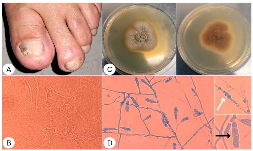
A 49-year-old female patient reports dysmorphic nail lesions of four years of evolution. On physical examination, the lesions extended to the toes of both feet, predominantly in the first toe of the left foot, which presented mild inflammatory changes in the soft tissue of the distal phalanx, accompanied by melanonychia, xantonychia, without paronychia, and with a thickened nail bed. Distal subungual onychomycosis was diagnosed. The nail of the left foot's first toe was scraped to collect scales, which were then clarified with 20% potassium hydroxide (KOH) for 30 min and were later examined under a microscope. Additionally, the samples were cultured in Sabouraud dextrose agar with chloramphenicol and cycloheximide (Liofilchem, Teramo, Italy) and incubated for 15 days at 28–30 °C. In the direct examination, thin and septate hyphae were observed. The culture showed colonies with a beige velvety appearance after 15 days of incubation. In the microscopic analysis, septate hyaline hyphae were observed, with abundant club-shaped macroconidia, thick walls divided by 2 to 6 septa, and without microconidia. The observed cellular and colonial morphology was compatible with Epidermophyton floccosum (Figure 1).
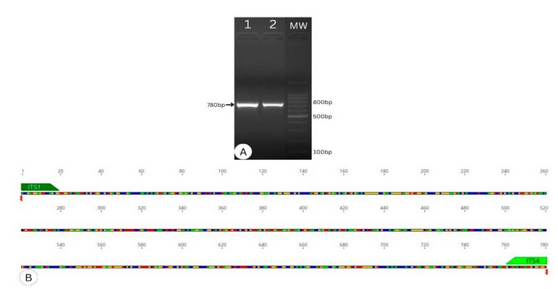
Molecular identification of the fungus was performed by amplifying and sequencing the ribosomal internal transcribed spacer region according to previously published 1, 2 (Figure 2). A 684 nucleotide sequence was edited using the Geneious prime® 2023.1.2 software. BLAST analysis confirmed the identity of E. floccosum. The sequence was deposited in GenBank under the accession number OR119955.

Figure 1. (A) Clinical characteristics of Epidermophyton floccosum-induced tinea unguium. (B) Direct examination with 20% KOH, where septate hyaline hyphae are observed. (C) Colonial morphology of E. floccosum on potato dextrose agar after 15 days of incubation at 28–30 °C. On the right side of Figure 1.C, the generation of yellow pigment is visible. In contrast, the colonies on the left side of the figure have a velvety crateriform appearance and are beige. (D) Microcultures of E. floccosum stained with lactophenol cotton blue. The white arrow shows the characteristic chlamydoconidium. The black arrow shows a characteristic club-shaped macroconidium.

Figure 2. (A) Agarose gel electrophoresis. Lanes 1 and 2 show a 780 bp band due to the amplification of the internal transcribed spacer (ITS) region. The 100 bp molecular weight (MW) marker is shown in the third lane. (B) In silico analysis depicting the target site of primers ITS1 and ITS4 to the ITS region and the expected amplicon size.
DISCUSSION
Dermatophytosis or ringworm is a fungal infection that affects the skin, hair and nails, with a prevalence of approximately 25% worldwide 3, 4. However, the prevalence of dermatophytes can range from 40 to 60% in some Asian and African nations(5, 6). Ringworms are caused by keratinophilic fungi known as dermatophytes 3, 4. Currently, it is known that at least nine genera are associated with dermatophytosis: Trichophyton, Microsporum, Epidermophyton, Lophophyton, Paraphyton, Nannizzia, Arthroderma, Ctenomyces and Guarromyces 3, 7, 8. Tinea unguium, often known as onychomycosis, is one of the most significant dermatophytoses. Onychomycosis is believed to be responsible for up to 50% of nail diseases, and its prevalence is estimated to be 10% worldwide(9). Onychomycosis usually occurs chronically and causes discoloration, onycholysis, and thickening of the nail plate9. It occurs most frequently in the toenails, and the most affected area is usually the nail of the first finger, where it can affect any component of the nail anatomy, including the nail bed, nail matrix, and nail plate.8, 10
Advanced age, diabetes mellitus, immunosuppression, especially that brought on by the Human Immunodeficiency Virus (HIV), tinea pedis, hyperhidrosis, obesity, peripheral vascular disease, and venous insufficiency are the key risk factors that affect the development of onychomycosis 10, 11.
Onychomycosis can be treated pharmacologically using three distinct approaches, including topical treatment, which uses formulations designed to be administered to the skin around and beneath the nails as well as the nails themselves. These drugs contain antifungals as an active ingredient, such as 8% ciclopirox, 5% amorolfine, and 10% efinaconazole. Oral antifungals such as terbinafine, itraconazole, and fluconazole are a second treatment choice. Among oral antifungals, terbinafine monotherapy with continuous doses of 250 mg remains the first-line treatment for these infections. The third treatment option is combination therapy, which combines topical and oral antifungals 9.
One of the agents that cause onychomycosis is the genus Epidermophyton, which was originally reported in 1907 12. Currently, E. floccosum is the only representative species of its genus 12. This fungus has been described as anthropophilic and has a very uneven distribution across the globe 4, 12. Less than 1% of dermatophytosis is caused by E. floccosum in the United States 13; however, over 14% of dermatophytoses recorded in Canada are caused by E. floccosum 13. According to reports, this fungus is responsible for between 1 to 15% of cases of dermatophytosis on the African continent, with Morocco and the Ivory Coast exhibiting the highest number of cases 6. In Europe, Ireland reported E. floccosum as the fourth causative agent of Tinea unguium, with a prevalence close to 15% 14, while in Greece, the prevalence of this fungus was 2.5%, which represented just a single case of Tinea unguium 15. However, only 0.3% of cases of dermatophytosis were caused by E. floccosum in Denmark.16.
In the Latin American region, some studies report that in Brazil, Mexico, and part of Central America, the presence of this fungus is 1% 17, 18. According to reports, E. floccosum cases comprised 1.2% of all dermatophytosis cases in Chile during the 1980s. However, during the 1990s, there was an almost 50% decrease in prevalence. By 2000, this prevalence had further decreased to less than 0.3% of all dermatophytosis cases 19.
CONCLUSION
This report presents the case of a 49-year-old female patient with Tinea unguium caused by Epidermophyton floccosum. There were no severe medical issues or underlying diseases that the patient faced. The extended wearing of closed shoes was the most significant risk factor found. Additionally, no similar occurrences in their families were reported. This clinical image is precious for educational purposes, as it can contribute to the knowledge and better understanding of dermatophytoses and promote learning in health area personnel. We believe this description would contribute to broadening our understanding of the epidemiology of dermatophytosis, particularly those caused by Epidermophyton floccosum. This is the first molecular characterization of Epidermophyton floccosum as an etiologic agent of Tinea unguium in Honduras.
Acknowledgments
None
Conflict of Interest Statement
The authors have no conflicts of interest to declare.
Funding
This research received no external funding. The experiments were conducted with the resources provided by the Genetic Research Center and the National University of Honduras School of Microbiology.
Consent
We have obtained the patient's consent in written form to publish the case report.
REFERENCES
1. Ortiz B, Enríquez L, Mejía K, Yanez Y, Sorto Y, Guzman S, et al. Molecular characterization of endophytic fungi from pine (Pinus oocarpa) in Honduras. Revis Bionatura 2022; 7 (3) 13. s Note: Bionatura stays neutral with regard to jurisdictional claims in …; 2015.
2. Ortiz B, Laínez-Arteaga I, Galindo-Morales C, Acevedo-Almendárez L, Aguilar K, Valladares D, et al. First molecular identification of three clinical isolates of fungi causing mucormycosis in Honduras. Infectious Disease Reports. 2022;14(2):258-65.
3. Martinez-Rossi NM, Peres NTA, Bitencourt TA, Martins MP, Rossi A. State-of-the-Art Dermatophyte Infections: Epidemiology Aspects, Pathophysiology, and Resistance Mechanisms. J Fungi (Basel). 2021;7(8).
4. Arenas Guzmán R. Micología médica ilustrada. 2014.
5. Verma SB, Panda S, Nenoff P, Singal A, Rudramurthy SM, Uhrlass S, et al. The unprecedented epidemic-like scenario of dermatophytosis in India: I. Epidemiology, risk factors and clinical features. Indian journal of dermatology, venereology and leprology. 2021;87(2):154-75.
6. Coulibaly O, L’Ollivier C, Piarroux R, Ranque S. Epidemiology of human dermatophytoses in Africa. Medical mycology. 2018;56(2):145-61.
7. de Hoog GS, Dukik K, Monod M, Packeu A, Stubbe D, Hendrickx M, et al. Toward a novel multilocus phylogenetic taxonomy for the dermatophytes. Mycopathologia. 2017;182:5-31.
8. Chanyachailert P, Leeyaphan C, Bunyaratavej S. Cutaneous Fungal Infections Caused by Dermatophytes and Non-Dermatophytes: An Updated Comprehensive Review of Epidemiology, Clinical Presentations, and Diagnostic Testing. Journal of Fungi. 2023;9(6):669.
9. Maskan Bermudez N, Rodríguez-Tamez G, Perez S, Tosti A. Onychomycosis: Old and New. Journal of Fungi. 2023;9(5):559.
10. Lipner SR, Scher RK. Onychomycosis: Clinical overview and diagnosis. Journal of the American Academy of Dermatology. 2019;80(4):835-51.
11. Albucker SJ, Falotico JM, Choo Z-N, Matushansky JT, Lipner SR. Risk Factors and Treatment Trends for Onychomycosis: A Case–Control Study of Onychomycosis Patients in the All of Us Research Program. Journal of Fungi. 2023;9(7):712.
12. Bonifaz Trujillo JA. Micología médica básica: Mc Graw Hill; 2010.
13. Ghannoum M, Hajjeh R, Scher R, Konnikov N, Gupta A, Summerbell R, et al. A large-scale North American study of fungal isolates from nails: the frequency of onychomycosis, fungal distribution, and antifungal susceptibility patterns. Journal of the American Academy of Dermatology. 2000;43(4):641-8.
14. Powell J, Porter E, Field S, O'Connell NH, Carty K, Dunne CP. Epidemiology of dermatomycoses and onychomycoses in Ireland (2001–2020): A single‐institution review. Mycoses. 2022;65(7):770-9.
15. Maraki S. Epidemiology of dermatophytoses in Crete, Greece, between 2004 and 2010. Giornale Italiano di Dermatologia e Venereologia: Organo Ufficiale, Societa Italiana di Dermatologia e Sifilografia. 2012;147(3):315-9.
16. Saunte DM, Svejgaard EL, Hædersdal M, Frimodt-Møller N, Jensen AM, Arendrup MC. Laboratory-based survey of dermatophyte infections in Denmark over a 10-year period. Acta dermato-venereologica. 2008;88(6):614-6.
17. de Oliveira Pereira F, Gomes SM, Lima da Silva S, Paula de Castro Teixeira A, Lima IO. The prevalence of dermatophytoses in Brazil: a systematic review. Journal of Medical Microbiology. 2021;70(3):001321.
18. Pérez-Rodríguez A, Duarte-Escalante E, Frías-De-León MG, Acosta Altamirano G, Meraz-Ríos B, Martínez-Herrera E, et al. Phenotypic and Genotypic Identification of Dermatophytes from Mexico and Central American Countries. Journal of Fungi. 2023;9(4):462.
19. Cruz R, Carvajal L. Frecuencia de Epidermophyton floccosum en dermatofitos aislados en un laboratorio de la Región de Valparaíso, Chile. Período 1980-2010. Revista chilena de infectología. 2018;35(3):262-5.
Received: 20 June 2023/ Accepted: 25 August 2023 / Published:15 September 2023
Citation: Zúniga B, Arita-Ramos M, Acevedo-AlmendárezL, García-Chávez J, Ponce-Mejía D, Fontecha G, Ortiz B. Tinea unguium caused by Epidermophyton floccosum. Revis Bionatura 2023;8 (3) 36. http://dx.doi.org/10.21931/RB/2023.08.03.36
