2023.08.03.80
Files > Volume 8 > Vol 8 No 3 2023
Histological study of liver in guinea pig Cavia porcellus ( Linnaeus , 1758) in Iraq.

Noor Hussein Yousif 1* , Hind Dyia Hadi 2 *, Hiba Mohammed Jihad 3*
*Iraq Natural History Research Center &Museum \ University of Baghdad
1 E mail: [email protected]
3 E mail: [email protected]
Corresponding author: [email protected] , Mobil-No. :+9647737157635
Available from: http://dx.doi.org/10.21931/RB/2023.08.03.80
ABSTRACT
The liver is one of the most prominent glands in the digestive system. It crosses vital organs with multiple functions, including the secretion of enzymes, digestion of fats, and secretion of bile. Through histological studies and those interested in them, the tissue structure of the liver is of interest to researchers, and the four samples of Guinea pig Cavia porcellus were taken in this study to know the histological structure and compare it with rodents in particular. and other animals in general. The results of the liver parenchyma were comparable to those of the studied mammals in periods of hepatocytes, Kupffer cells, and sinusoids surrounding the central vein, blood supply, and bile ducts.
He did not record differences, even if they were minor, commensurate with the size of the animal and the environment in which it lives.
Key word : Guinea pig, Cavia porcellus, Liver, Hepatocyte , Kupffer cell, Iraq
INTRODUCTION
Rodents are classified as one of the most essential mammals used in laboratories, conducting tests, and scientific research1. The liver is a complex gland covered by peritoneal epithelium with rows of cells resting on basal connective tissue containing collagen fibrils 14,15. During embryonic development, the liver develops from the primary alimentary gut, which produces lobes containing hepatocytes separated by vascular channels that extend into sinusoids later. After being absorbed from the stomach, food will pass into the liver directly through the hepatic portal vein2. In previous studies, the liver was known to be one of the largest organs in the mammalian body. It is a lobed organ into several lobes anatomically up to 5 lobes. Histologically, histology is essential to differentiate between normal cells and cells that change according to diseases 3,4; The liver is a connective tissue and blood vessels surrounded by a fibrous capsule5.
The liver is an endocrine gland; its large size makes it the largest gland in the body within the digestive system. It contains two types of secretion: the external secretion, represented by the bile and the internal secretion, characterized by the secretion of protein and amino acids. It controls metabolic activities and plays an essential role in eliminating toxic and harmful substances inside the body 6.
The primary tissue of the liver parenchyma is a collection of lobules and reticular fibers, interspersed with neural and hematopoietic synapses and characterized by the presence of Kupffer cells 7.8.
Consecutive studies conducted on the liver showed that factors affecting the shape of liver cells, such as stress, tension, environment, and embryonic development, through the adaptation of animals to their environment2,9. This study aims to determine whether there are histological differences in the liver parenchyma in animal guinea pigs and previously studied animals.
MATERIAL AND METHOD
Liver tissue samples were taken from adult male guinea pigs and the samples were carefully cleaned and fixed with 10% diluted formalin for 48 h. After fixation, it was placed in Bouin's solution for 15 hours, then the sample was passed through a series of dilutions of ethyl alcohol solution (70%, 80%, 90%, and 100%) for two consecutive hours for each dilution. Other tissue incisions were made after which the sample was stained (hematoxylin and Eosin and Masson Trichome stain10, 11. Tissue sections were photographed using a digital camera.
RESULT
Histological results of the microscopic examination for the current study present the liver's soft tissue surrounded by a thin capsule composed of dense regular connective tissue fibers and liver parenchyma consisting of the reticular fiber and hepatocytes (Fig,1A&4). Liver is composed of lobules which consist of a central vein It divides the lobules trabeculae extending from the capsule wall of the liver into beneath the lobules surrounded the connective tissue distinguishing the lobules from the adjacent lobules (Fig,1A and C). Hepatocytes clearly arranged radials around the vein present between them the sinusoids(Fig,2 and 3). Hepatocytes are evident polygonal or irregular cells with a central nucleus in some cells with two nuclei arrest on the base membrane The cytoplasm of eosinophils in hepatocytes has few vacuoles containing fine grains. The lobules are composed of two raw hepatocytes with middle central vein (Fig,3), a portal region (Fig,1B) organized around the portal area containing a branch hepatic vein, a branch of the hepatic artery, a branch of lymphatic and a canal of the bile duct, as we notice the presence of defensive Kupffer cells that protrude clearly(Fig,3) beneath sinusoids circular cells with dark rounded nuclei but not directly attached to the basement membrane. Sinusoids are filled with numerous erythrocytes, and lined with endothelial cells and are separated from hepatic cells raw by the space of intersinusoids(Fig,4).
The guinea pig tissue of the liver in the current study did not differ from the liver in the other mammals.
The portal area was clear lumen-wide. the hepatic vein was lined with simple squamous epithelium. the hepatic artery linings with smooth muscle fiber and very clear inner and outer tunica(Fig,1B).
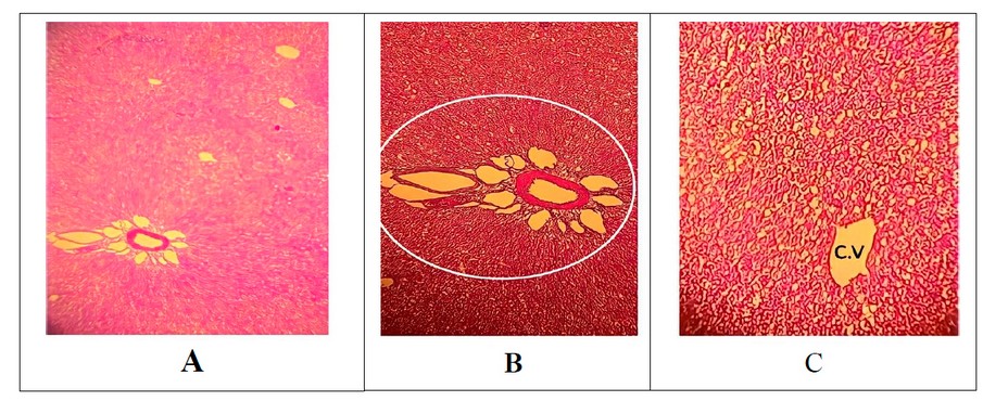
Figure 1. section through liver shows: A) the liver parenchyma(portal area ,radial hepatocyte around central vein . H&E stain 10x. B)Portal area (branch hepatic vein ,branch of hepatic artery , branch lymphatic and chanal of bile duct) H&E stain 40x. C) central vein. H&E stain 10x.
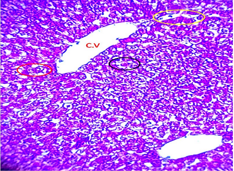
Figure 2. section through the liver shows the central vein, hepatocyte (red circle), sinusoid (yellow ring)and kuffer cell (black circle ). Masson trichrome stain 100x.
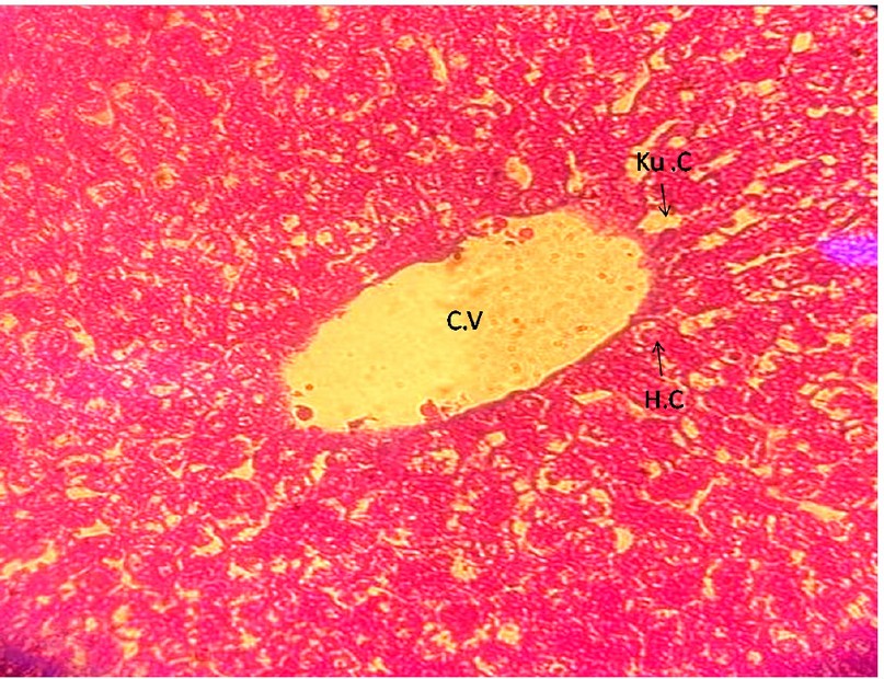
Figure 3. section through liver shows: central vein , Hepatocyte (H.C) , and kuffer cell (Ku.C ) .H&E stain 100x.
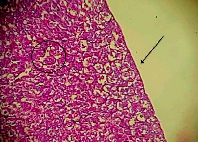
Figure 4. section liver shows: capsule (black arrow) and sinusoid (black circle . Masson trichome stain 100x..
DISCUSSION
Through the studies presented by12 ,the Egyptian rodents13 European Bison14 , and the African palm squirrel15 . It agreed with the current study that the tissues are interconnected and divided into lobules separated from each other by trabeculae. The current study is similar to that16,17 in that the liver is composed of the liver parenchyma which included the central vein around which the liver cells lined up in two rows or one row separated by hepatic sinusoids. Hepatocytes were radially raised around the central vein, which branched into branches, then returned and met to encircle the hepatic vein that occupies the middle of the liver lobe and the hepatic sinuses that are lined by endothelial cells, with the protrusion of defensive Kupffer cells with one distinct dark nucleus into the lumen With the presence of binucleated liver cells that are fully prepared for reported, in the event of compensation, if any, the study was consistent with humans, camels, cows, goats and Sheep, rodents, and African Palm Squirrel 13,15,17,18 . The presence of hepatic sinusoidal cells and Kupffer cells in few numbers that are easily identifiable was consistent with other studies camel, Cows, and African Palm Squirrel15,18 .
When comparing the current study with9 shows regular-shaped liver cells separated by hepatic sinusoids, red blood cells appear with Kupffer cells protruding with the endothelial cells of the hepatic sinusoids, The physiological function of Kupffer cells is a defensive function represented by devouring foreign bodies and toxic substances that come from the portal artery consistent with previous studies squirrel and Iraqi mammals 2,9. Compared to large mammals, the liver of H. javanicus and S. carolinensis was similar to the current study in terms of the capsule that surrounds the liver parenchyma with fibers and hepatocytes, and this was consistent with the study of both humans and wild ruminants 19,20.
Hepatocytes are physiologically responsible for all liver functions, and the difference in this shape is due to genetic factors, lifestyles, and the environment that affects the animals' life, including all animals16.
Cytoplasm shape in the liver differed in studies that may be attributed to the function of the liver in storing glycogen and lipids. The examination corresponds to the 21.
CONCLUSIONS
Histological liver studies show no differences between mammals, as they carry within themselves the same collagenous connective tissue of the parenchyma, hepatocytes, and sinusoids, meaning that the difference may arise due to environmental conditions or stress. This is what resulted in the current study. It is histologically identical to all studies conducted on the liver in different animals. Statistical and quantitative analyses of liver physiology are recommended. Perhaps it would also be best to use electron microscopy to describe the liver parenchyma in it.
ACKNOWLEDGMENTS
This study is supported by the Research Program of Iraq Natural History Research Center and Museum \ University of Baghdad, Iraq. My thanks to my teachers and colleagues in the scientific process.
DISCLOSURE OF CONFLICT OF INTEREST
There was no conflict of interest in this study.
STATEMENT OF ETHICAL APPROVAL
All operations and conclusions were executed under the knowledge and supervision of the laboratories of the Iraq Natural History Research Center and Museum\ University of Baghdad.
REFERENCES
1- Nzalak J. O,Onyeanusi B.I, Salami and S.O. Macrometric study of the digestive system of the African giant rat (Cricetomys gambianus, Waterhouse 1840). Eur. J. Anat., 2012, 16 (2):113-8.
2- Yousif, N. H. Histological study of liver for squirrel (Sciurus anomalus)(Güldenstädt, 1785) in Iraq. GSC Biological and Pharmaceutical Sciences, 2022, 20(1), 091-094.
3- Copenhaver, W. M. Bailey's Textbook of Histology. The Williams and Wilkins Company, Baltimore, 1964.
4- Banks, W. J. Applied Veterinary Histology. Third Edition, Mosby Books, USA. ,1993.
5- Petcoff G M, Díaz A O, Escalante A H and Goldemberg A L, Histology of the liver of Oligosarcusjenynsii (Ostariophysi, Characidae) from Los Padres Lake, Argentina. Iheringia, Sér.Zool. 2006, 96 (2), 205-208.
6- Akiyoshi, H. & Inoue, A. Comparative Histological Study of Teleost Livers in Relation to Phylogeny. Zoolog. Sci., 2004, 21 (8):841-50.
7 -NAFADY, Allam AM, AWADALLA, Eatemad A. Journal of Advanced Trends in Basic and Applied Science Vol. 1, No. 2: 148-158, 2017.
8- Carollo, V.; Giancamillo, D. A.; Vitari, F.; Schneider, R. & Domeneghini, C. Immunohistochemical Aspects of Ito and Kupffer Cells in the Liver of Domesticated and Wild Ruminants. Open J. Vet. Sci. 2012, 2 (3):129-36.
9- AL-Aamery, Rana Alaa, et al. morphological description and comparative histological study of the liver in two iraqi mammals: weasel (herpestes javanicus) and eastern gray squirrel (sciurus carolinensis). Biochem. Cell. Arch. 2020 , 20(1): 167-170.
10-Culling, C. F. A., Allison, R. T., and Barr, W. T. Cellular pathology technique. Elsevier, 2014.
11-Luna,L." Manual of Histological Staining Methods the Armed Forced Institute of Pathology "3rdEd.American Registry of Pathology.New York. 1968: 76-98.
12- Rogers A B and Dintzis R Z . Hepatobiliary system in Comparative anatomy and histology: a mouse, rat, and human atlas (eds: Treuting P M, Dintzis S M and Montine K S) 2nd edn., Academic press an imprint of Elsevier, London,U.K. 2018: 229-240.
13- EL-SALKH, Boshra A., et al. Anatomical, Histological and histochemical studies on some organs of true desert rodents in the Egyptian habitats. Egypt J Hosp Med, 2008, 33(1): 578-306.
14- Prunescu P P, Prunescu P, Krasinska M and Krasinsk Z, A Liver histological structure in adult European Bison. Bison bonasus (Linnaeus, 1758). Folia Morphol. 2002, 61 (3), 137–142.
15- Ikpegbu E, Nlebedum U C, Nnadozie O and Agbakwuru I O) The Liver Micromorphology of the African Palm Squirrel Epix erusebii. Int. J. Morphol. 2014, 32 (1), 241-244.
16- Frappier B L . Digestive system in Dellmanns text book of veterinary histology (eds. Eurell J A and Frappier B L). 6th edn., Black well publishing. 2006: 201-206.
17 - Madhan K E and Raju S. Comparative Histology of human and Cow, Goat and Sheep liver. Journal of Surgical Academia .2014, 4(1):10-13.
18- Alshamarry H A, Sucar D K and Taha T J Comparative Histological study for The Iraqi. Camels (Camelus dromedarius) liver & Cows (Bos indicus). Journal of Thi-Qar Science 2010, 2 (3), 39-48.
19- Madhu D . Morphological studies on the visceral organs of wild ruminants. M.V.Sc. Thesis. Department of Veterinary Anatomy and Histology, Veterinary College, Sciences University, BIDAR, 2013: 129.
20- Madhan K E and Raju S Comparative Histology of human and Cow, Goat and Sheep liver. Journal of Surgical Academia 2014, 4 (1), 10-13.
21- Kareem S . Histological study of the native rabbit liver. Al- Qadisiyah Journal of Veterinary Medicine Sciences ,2013,12(2), 69-74.
Received: 25 June 2023/ Accepted: 26 August 2023 / Published:15 September 2023
Citation: Yousif N H, Hadi H D, Jihad H M. Histological study of liver in guinea pig Cavia porcellus ( Linnaeus , 1758) in Iraq.Revis Bionatura 2023;8 (3) 80. http://dx.doi.org/10.21931/RB/2023.08.03.80