2023.08.03.118
Files > Volume 8 > Vol 8 No 3 2023 > Diyala 3
Identification and Antifungal susceptibility of Rhodotorula mucilaginosa isolated from women patients in Erbil City-Iraq Kurdistan

Nareen Qadr FaqeAbdulla*
Biology Department, College of Sciences, University of Salahaddin, Erbil, Kurdistan Region, Iraq. [email protected]
* Correspondence: [email protected]
Available from. http://dx.doi.org/10.21931/RB/2023.08.03.118
ABSTRACT
Rhodotorula is emerging as an essential cause of nosocomial and opportunistic infections. This is the first study on Cervical-Vulvovaginal yeast infections aimed to find the prevalence of Rhodotorula mucilaginosa as the causative agent of Cervical-Vulvovaginal infection in women in Erbil City. For identification methods, API 20C AUX system, CHROMagar and Multiplex PCR amplification of universal primer confirmed by DNA sequencing using the ITS1 and ITS4 universal primer. Sequence data of Rhodotorula mucilaginosa provided GenBank accession number OQ568163 for nucleotide sequence. According to Congo red agar (CRA) and molecular method for amplifying the ALS1 and HWP1 genes, the isolates produced biofilms. Furthermore, evaluating the antifungal susceptibilities of Rhodotorula mucilaginosa by disc diffusion method revealed that the isolates were sensitive to all tested antifungal discs: Econazole (20mm), Ketoconazole (22mm), Nystatin (24mm), except Miconazole (9mm), which had a little effect on inhibition zone. Agar Well Diffusion Method showed that the isolates were resistant to all tested antifungal drugs: Griseofulvin (4mm), Itraconazole (0mm), Ketoconazole (6mm), and Nystatin (6mm), except Fluconazole (24mm), which affected inhibition zone.
Keywords: Rhodotorula mucilaginosa, Econazole, Fluconazole, CHROM Agar, biofilm
INTRODUCTION
In regular practice, vulvovaginitis is an issue that is frequently encountered. Vaginal discharge and discomfort are the typical first signs of it. To avoid missing the less prevalent causes, a comprehensive evaluation is necessary: 1. Rhodotorula are common environmental yeasts once considered non-pathogenic. However, over the past three decades, various species of this yeast have established themselves in people as pathogens, leading to systemic illnesses in the immunocompromised N2. Treatment of such novel infections requires knowledge of the Rhodotorula strains' susceptibility to commonly used antifungal medications3. The yeasts in the genus Rhodotorula are saprophytic. R. mucilaginosa, R. glutinis, and R. minuta are species of the genus, but none have yet been linked to human disease4. Recent research has identified the pigmented yeast Rhodotorula spp. as an emerging opportunistic pathogen that can colonize and infect immunocompromised patients and cause oral ulcers, aortic homograft endocarditis, meningitis, and peritonitis dermatitis 5-7. Blood cultures can become contaminated, and Rhodotorula mucilaginosa may be on the skin. Due to the multiresistant profile, fungemia caused by R. mucilaginosa is a rare clinical entity that requires risk factors 8. Regarding the epidemiology, risk factors, and consequences of Rhodotorula-related mycoses in people, very little scientific information is available 9,10. The majority of these systemic infections are treated using antifungal agents such as amphotericin B and azoles viz. ketoconazole, fluconazole etc. 11. The objective of this study was to isolate and identify species of Rhodotorula mucilaginosa from cervical-vulvovaginal samples of women patients by using the conventional and molecular-based method and its identification was confirmed by DNA sequencing using universal primers, ITS1 and ITS4 and detection of biofilm virulence gene, ALS1 and HWP1, then determine the efficacy of different antifungal discs and drugs against it.
MATERIALS AND METHODS
Collection and Isolation of samples
Fifty strains were collected from Cervical-Vulvovaginal patients' samples, including signs and symptoms of VVC assessed during the gynecological exam at different hospitals. Prior to the test, the positive isolate samples were cultivated on Sabouraud᾽s dextrose agar medium, with the addition of antibiotics to prevent the growth of bacteria. After 3–5 days of incubation at 37ºC, cultures were separated into positive (yeast growth) or negative cultures (no yeast growth). Using this approach, we isolated and determined the number of yeast colonies12.
Phenotypic identification of yeast
Microscopic examination: Each sample was stained with lactophenol cotton blue and inspected under a microscope (40X).
Chromogenic medium (CHROM Agar Candida): After incubation (48hr at 37°C), all positive cultures were cultured on CHROM Agar (BioMérieux, France); yeast identification was performed based on colony color.
Biotyping identification: The yeast isolates were biochemically identified using API 20C AUX strips. A single colony from a young yeast culture obtained from an SDA plate was submerged in an API 20C suspension tube. The strips were incubated for (24, 48, and 72) hr at 37°C after being filled with the suspension from the API 20C medium tube in the cupules of the strip. Following incubation, cupule turbidity was measured recorded, and a profile number was created13.
Molecular identification via amplification of ITS region
DNA extraction and PCR amplification: The yeast isolates were grown on SDA at 37ºC for 48 hours, and then DNA was extracted using a Genomic DNA isolation kit (Fungi/Yeast Genomic DNA isolation kit. Norgen Biotek/Canada), depending on the manufacturer's instructions. The internal spacer region was amplified by using universal primers14. A total of 25µl PCR master mix reaction volume was performed containing 3 µl of genomic DNA, 12.5µl of 2X GoTaqGreen Master Mix (Promega/USA), and 1µl was added for each of the forward and reverse primer for both ITS1 and ITS4, forward (ITS1, F'5-TCC GTA GGT GAA CCT GCG G-´3), reverse (ITS4, R-5' TCC TCC GCT TAT TGA TAT GC-´3) primer then the mixture was completed by adding 7.5µl of nuclease-free water. The thermal cycles were programmed as follows: initial denaturation cycles at 95°C for 5 min., followed by 35 cycles at 94°C for 1 min., 55°C for 1min., 72°C for 1min. and a last extension at 72°C for 7 minutes. The 505bp of PCR products were confirmed by using 2% agarose gel electrophoresis in 1XTBE buffer, and PCR product of yeast isolates was sent to (Macrogen/South Korea) for sequencing.
Sanger Sequencing Technique for Fungal Strain Identification: Integrated DNA technologies/USA provided oligonucleotide primers for polymerase chain reaction. The internal transcribed spacer regions ITS1 and ITS4 were amplified using nonspecific primers. All Fungal isolates' targeted DNA was effectively amplified, with PCR amplicon sizes ranging from 500 to 800bp15.
Genetic analyzer 3500 (Macrogen/South Korea) was used to test the amplified amplicons of ITS isolates. The fasta file sequences were modified, aligned, and submitted to GeneBank to obtain an Accession number, after which they were compared to other reference strains from the different sequences.
Detection of biofilm formation by Congo red method (CRA):
It is an alternative method for biofilm formation described by Freeman et al., 16. A positive result was indicated by black colonies with a dry crystalline consistency. A non-biofilm producer usually remains pink. The experiments were performed in triplicate and repeated three times.
Detection genes associated with biofilm formation:
Amplification of ALS1 and HWP1 genes: PCR amplification was carried out by using ALS1 primers Forward (F'5-GAC TAG TGA ACC AAC AAA TAC CAG A -' 3) and Reverse (R-5'CCA GAA GAA ACA GCA GGT GA-´3) and HWP1 primers Forward (F'5- ATG ACT CCA GCT GGT TC), Reverse (R-5'TAG ATC AAG AAT GCA GC-´3). A total of 25 PCR master mix reaction volumes was performed containing 3µl of genomic DNA, 12.5 µl of 2X GoTaqGreen, Master Mix (Promega/ USA) and 1µl was added for each of the forward and reverse primer for both genes and volume completed with 5.5µl of DNase, RNase free water. The PCR was conducted as follows: 1 cycle of 94°C for 4min., followed by 35 cycles of 94°C for 30s, 52°C for 1min., and 72°C for 2min. A final extension cycle was performed at 72°C for 5 minutes. The PCR products were separated on 2% agarose. The 318bp and 572bp mean the amplification was successfully done for ALS1 and HWP1 genes17.
Antifungal susceptibility test
Antifungal disc by Disc Diffusion Method (DDM):
This is the current reference method recommended by the National Committee for Clinical Laboratory Standards (NCCLS). The Kirby-Bauer disk diffusion method evaluated yeast isolates in vitro for antifungal drug susceptibility. The disks used in the testing were Biorex diagnostics microbiology sensitivity discs manufactured in the U.K., mainly Econazole50mg, Ketoconazole50mg, Miconazole50mg and Nystatin100mg. It was performed on SDA agar supplemented with 8% glucose.
Antifungal drugs by Agar Well Diffusion Method (AWDM):
The antifungal drugs, including Fluconazole, Griseofulvin, Itraconazole, Ketoconazole, and Nystatin, were tested against Rhodotorula mucilaginosa. The stock solution was prepared for each drug, except for Itraconazole, which was dissolved using DMSO; other medicines' stock solutions were made by dissolving them in SDW. The inoculum of Rhodotorula mucilaginosa was prepared using 48hr fresh yeast cultures grown on SDB, and they were adjusted to (1x10⁶/ml) with a bright line hemocytometer (Hausser Scientific, Horsham, Pa)18. Briefly, 0.1mL (100µL) suspension of each isolate was spread over SDA; a sterile cork borer was used to punch 6mm diameter wells in the culture media. Then, 100µL of each drug was poured into each well until complete. The treated plate was incubated at 37°C for 24 hours. The diameters of inhibition zones were measured in millimeters using a ruler for each antifungal drug 19.20.
Statistical Analysis
The distribution of the data was analyzed according to the Shapiro-Wilk and Kolmogorov-Smirnov normality test. Then, the data were analyzed according to a one-way analysis of variance (ANOVA) followed by multiple comparisons of Tukey's test. For other obtained data, an unpaired t-test was applied using GraphPad Prism software (version 9). P< 0.05 is considered statistically significant.
RESULTS
Phenotypic identification: The identification of the samples isolated from cervical-vulvovaginal patients is phenotypically summarized in (Table 1). Nine non-duplicated Rhodotorula mucilaginosa isolates from Vulvovaginal candidiasis (VVC) patients in Erbil City-Iraqi Kurdistan. Identification was performed on SDA; the colony color glistening, smooth, mucoid, orange-pink colonies of Rhodotorula mucilaginosa. These isolates failed to produce germ tube in human serum after 3 hours of incubation at 37∘C. Identification was performed on CHROM Agar after incubation 48hr at 37˚C based on colony color. By using this method, the color of Rhodotorula mucilaginosa has not changed.
Table 1. Phenotypic characteristics of Rhodotorula mucilaginosa isolates
Molecular identification (PCR and sequences): The result of the molecular identification, by using ITS1 and ITS4 gene Amplification, was consistent with the phenotypic analysis (Figure 1). Isolated Rhodotorula shared the identical ITS sequence, which differs from others in one position within the 35 bp ITS2 signature sequence. The amplification products were submitted to sequencing analysis and showed 99% to 100% sequence identity with the Rhodotorula strains. Sequence data of Rhodotorula provided GenBank accession number OQ568163 for nucleotide sequence.

Figure 1. PCR amplification of Internal Transcribed spacer region. Lane L: DNA ladder 1kb, Lane 2: negative control 3: Correspond to Fungal ITS region amplification.
Biofilm formation: In this study, Congo red Agar was used for the detection of the ability of Rhodotorula mucilaginosa to form biofilm. It was revealed that all Rhodotorula mucilaginosa, isolated from women patients with cervical-vulvovaginitis were biofilm producers.
Detection of ALS1 and HWP1 genes associated with biofilm formation: The prevalence of the studied virulence genes in Rhodotorula mucilaginosa isolates was HWP1 and ALS1 using PCR. The gene HWP1 was found, but ALS1 was not found in Rhodotorula mucilaginosa. In the present study, biofilm formation was evaluated to see the reflection of gene presence on phenotype (Table 2, Figure 2).
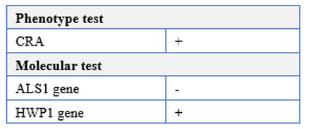
Table 2. Phenotypic and molecular test of Rhodotorula mucilaginosa for biofilm formation
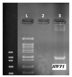
Figure 2. Agarose gel electrophoresis of ALS1 and HWP1 gene amplification (PCR products) for biofilm formation Rhodotorula mucilaginosa.
Susceptibility testing of antifungal drugs
In this study, all the isolates of Rhodotorula mucilaginosa were studied against antifungal disc and drugs based on the zone of inhibition of growth. Disk diffusion test was performed on SDA agar; the ANOVA test showed significant differences in the sensitivity of Rhodotorula mucilaginosa against antifungal discs. It was reported as sensitive for each of the tested antifungal's discs: Econazole50mg (20mm), ketoconazole50mg (22mm) and Nystatin100mg (24mm), while resistance for Miconazole50mg (9mm), which had a little effect on inhibition zone. The zone of inhibition showing the interpretive categories by the disk diffusion technique is presented in (Table 3, Figures 3 and 4). For Antifungal drugs, the ANOVA test showed significant differences in the sensitivity of Rhodotorula mucilaginosa, including Fluconazole, Griseofulvin, Itraconazole, Ketoconazole, and Nystatin. It revealed that all isolates were resistant to Griseofulvin (4mm), Itraconazole (0mm), Ketoconazole (6mm), and Nystatin (6mm), which did not affect inhibition; nevertheless, tested isolates were sensitive to Fluconazole (24mm) which affected inhibition zone (Table4, Figure5 and 6).
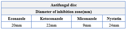
Table 3. Susceptibility testing of Rhodotorula mucilaginosa isolates against the antifungal disc
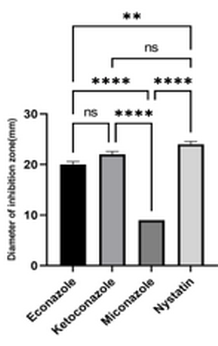
Figure 3. Quantitative measurement of inhibition zone for Rhodotorula mucilaginosa to antifungal discs by DDM. Data are presented as mean ±SE of three biological replicas. All data were significant at P<0.0001(***).
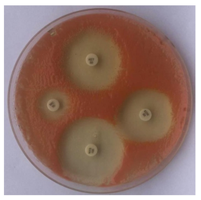
Figure 4. Disc diffusion method with antifungal azoles: Econazole, Ketoconazole, Miconazole and Nystatin
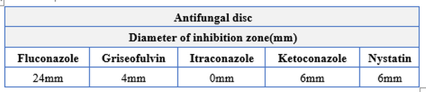
Table 4. Susceptibility testing of Rhodotorula mucilaginosa isolates against antifungal disc
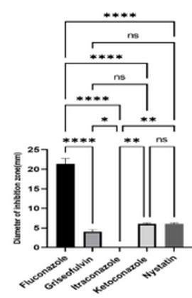
Figure 5. Quantitative measurement of inhibition zone for Rhodotorula mucilaginosa to antifungal drugs by AWDM. Data are presented as mean ±SE of three biological replicas. All data were significant at P<0.0001(***).
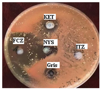
Figure 6. Agar diffusion method with antifungal azoles: FCZ, Ketoconazole KET, and Nystatin NYS Gris, ITZ.
DISCUSSION
An understanding of the role of yeasts in the environment has been uncertain because population size and diversity estimates have often been based on species identifications that were determined from a limited number of phenotypic characteristics. The widespread use of DNA-based species identification now enables a precise evaluation of species in various settings. However, because specific taxa are polyphyletic, there are still classification issues. As a result, it is still challenging to identify yeasts, estimate their genus-level diversity, and assign genera to higher taxonomic orders21.
Historically, Rhodotorula species were regarded as contaminants, but during the past two decades, they have gradually come to be understood as human infections. Although less virulent than Trichosporon or Candida, reports have revealed that Rhodotorula can produce severe, sometimes deadly, invasive infections 22.
Almeida et al., described 25 cases of Rhodotorula spp. Isolated from blood cultures at a Brazilian tertiary teaching hospital and investigated the in vitro activity of four antifungal drugs using a standardized method. All strains were identified as R. mucilaginosa by conventional methods. Misidentification of the species was observed with the Vitek Yeast Biochemical Card and API 20C AUX systems; Fluconazole demonstrated good activity high for all strains23.
Rhodotorula mucilaginosa yeasts can be challenging to distinguish phenotypically 24. In order to accurately identify these opportunistic fungal infections for surveillance, molecular screening is essential. It is imperative to conduct more research to understand the mechanisms of R. mucilaginosa infection, mycoparasitism in the human oral cavity, and the numerous underlying factors that may raise the risk of angular cheilitis25.
Rhodotorula infections are uncommon, but given the expected evolution of the virulence factors, it is likely that Rhodotorula infections will become more common in the future, as shown by the increasing number of case reports in the microbiological literature26.27.
Using the disk diffusion technique, the antifungal drugs ketoconazole, nystatin, and Fluconazole were evaluated in Rhodotorula, which showed sensitivity to ketoconazole and nystatin. No antifungal sensitivity to Fluconazole has been reported (2). In homemade fermented rice water, Rhodotorula sp. was discovered utilizing phenotypic techniques, such as biochemical traits using API ID 32C and molecular techniques28.
Turchetti et al. isolated 23 yeast strains that could not be assigned to any known fungal taxa during ecological studies of yeast communities carried out in cold ecosystems in the Italian Alps, Svalbard (Norway, Arctic region), and Portugal; in particular, two of them were first identified as Rhodotorula sp.29.
CONCLUSIONS
This is the first identification of Rhodotorula mucilaginosa from cervical-vulvovaginal candidiasis of women patients in Erbil city, Iraqi Kurdistan. A conventional sequencing technique was used to identify the Rhodotorula mucilaginosa, which was a reliable, rapid, and cost-effective technique. Biofilm production and antifungal drug resistance were observed in Rhodotorula mucilaginosa. Biofilm production could act as one factor in reducing the antifungal agent's penetrability. The biofilm virulence gene ALS1 was undetected, while HWP1 was detected in isolated strains. Funding: We relied on ourselves in all aspects, as no research funding sources were reported. Informed Consent Statement: The patient's consent was made in writing through the term of informed consent after the human ethics committee of the University of Salahaddin-Erbil approved this study. Inclusion criteria were vaginal swabs from women with cervical-vulvovaginal candidiasis who tended to participate in the study. All procedures performed in this study involving women participants followed the rules on patient sampling and fully complied with the ethics committee.
Acknowledgments: We thank the patient for participating in the study. Our special thanks go to the Maternity Teaching Hospital at the gynecology clinic in Erbil City, Iraqi Kurdistan and the University of Salahaddin College of Science-Biology Department for providing the opportunity to carry out the work.
Conflicts of Interest: The authors declare no conflict of interest.
REFERENCES
1. Sheppard C. Treatment of vulvovaginitis. Aust Prescr. 2020;43(6):195-9.
2. Nur F, Ferdoush S, Shahid MS, Begum K. Phenotypic Identification and Antifungal Susceptibility of Two Rhodotorula Species Isolated from Dandruff Samples. Dhaka University Journal of Pharmaceutical Sciences. 2020;19(2):185-90.
3. Krzyściak P, Macura AB. Drug susceptibility of 64 strains of Rhodotorula sp. . Wiadomoœci Parazytologiczne. 2010;56(2):167–70.
4. Larone DH. Medically Important Fungi: A Guide to Identification. 3rd ed., ASM Press, Washington (D.C.), p. 274. 1995.
5. Fell J.W., Boekhout T, Fonseca A, Scorzetti G, Statzell-Tallman A. Biodiversity and systematics of basidiomycetous yeasts as determined by large-subunit rDNA D1/D2 domain sequence analysis. Int J Syst Evol Microbiol. 2000;50:1351-71.
6. Duggal S, Jain H, Tyagi A, Sharma A, Chugh T.D. Rhodotorula fungemia: two cases and a brief review. Med Mycol. 2011;49(8):879-82.
7. Means AD, Sisto K, Lichon V, Monaghan D, O'Keefe P, Tung R. Cutaneous rhodotorula treated with photodynamic therapy. Dermatol Surg 2012;38:1100-3.
8. Falces-Romero I, Cendejas-Bueno E, Romero-Gómez MP, García-Rodríguez J. Isolation of Rhodotorula mucilaginosa from blood cultures in a tertiary care hospital. Mycoses. 2018;61(1):35-9.
9. Tuon FF, Duboc de Almeida GM, Costa SF. Central venous catheter-associated fungemia due to Rhodotorula spp.–a systematic review. Med Mycol. 2007;45:441- 7.
10. Tuon FF, Costa SF. Rhodotorula infection. A systematic review of 128 cases from literature. Rev Iberoam Micol 2008;25:135-40.
11. Diekema DJ, Petroelje B, Messer SA, Hollis RJ, Pfaller MA. Activities of available and investigational antifungal agents against Rhodotorula species. J Clin Microbiol. 2005;43:476-8.
12. Babic M, Hukic M. Candida albicans and non-albicans species as etiological agent of vaginitis in pregnant and non-pregnant women. Bosnian journal of basic medical sciences. 2010;10(1):89-97.
13. Shinoda T, Kaufman L, Padhye AA. Comparative evaluation of the Iatron serological Candida check kit and the API 20C kit for identification of medically important Candida species. J Clin Microbiol. 1981;13(3):513-8.
14. Jafari Z, Motamedi M, Jalalizand N, Shokoohi GR, Charsizadeh A, Mirhendi H. Comparison of CHROMagar, polymerase chain reaction-restriction fragment length polymorphism, and polymerase chain reaction-fragment size for the identification of Candida species. Curr Med Mycol. 2017;3(3):10-5.
15. W Mansoor, S. S.; Al-Esawi, J. S. . .; Al-Falahi, M. N. Assessing The Efficiency Of Cement Kiln Dust For Heavy Metals Removal From Simulated Polluted Water. JLSAR 2023, 4, 45-52.
16. Othman Ghazi Najeeb Alani , Yassen Taha Abdul-Rahaman and Thafer Thabit Mohammed. Effect Of Vêo® Premium and Vitamin C Supplementation on Lipid Profile Before and During Pregnancy in Some Local Iraqi Ewes During Heat Stress. Iraqi Journal of Science.2021, Vol. 62, No. 7, pp: 2122-2130.
17. İNCİ M, ATALAY MA, ÖZER B, EVİRGEN Ö, DURAN N, MOTOR VK, et al. Investigations of ALS1 and HWP1 genes in clinical isolates of Candida albicans. Turkish Journal of Medical Sciences. 2013;43:125-30.
18. Aboualigalehdari E, Sadeghifard N, Taherikalani M, Zargoush Z, Tahmasebi Z, Badakhsh B, et al. Anti-biofilm Properties of Peganum harmala against Candida albicans. Osong Public Health Res Perspect. 2016;7(2):116-8.
19. S. M. Abdulateef, O. K. Atalla1, M. Q. A L-Ani, TH. T Mohammed, F M Abdulateef And O. M. Abdulmajeed. Impact of the electric shock on the embryonic development and physiological traits in chicks embryo. Indian Journal of Animal Sciences.2021, 90 (11): 1541–1545.
20. Srinivasan D, Nathan S, Suresh T, Lakshmana Perumalsamy P. Antimicrobial activity of certain Indian medicinal plants used in folkloric medicine. Journal of ethnopharmacology. 2001;74(3):217-20.
21. Takashima M, Sugita T, Van BH, Nakamura M, Endoh R, Ohkuma M. Taxonomic richness of yeasts in Japan within subtropical and cool temperate areas. PLoS ONE. 2012;7(11):e50784.
22. Merkur AB, Hodge WG. Rhodotorula rubra endophthalmitis in an HIV positive patient. . Br J Ophthalmol 2002;86:1444-5.
23. Almeida DD, Gisele M, Costa F, Silvia, Melhem, Marcia, et al. Rhodotorula spp. isolated from blood cultures: Clinical and microbiological aspects. Medical Mycology. 2008;46:547-56.
24. Libkind D, Sampaio J. Rhodotorula. In: Liu D, editor. Molecular detection of foodborne pathogens. Boca Raton: CRC Press. 2010:p. 603–18.
25. Ibraheem M W, Muhaimeed A R, Mohammed Th. T. Leg cuts from Awaasi lambs fed a diet with varying levels of Rhus coriaria L., Physical dissection and chemical composition. Revis Bionatura 2022;7(4) 4. http://dx.doi.org/10.21931/RB/2022.07.04.4.
26. Savini V, Sozio F, Catavitello C, Talia M, Manna A, Febbo F, et al. Femoral prosthesis infection by Rhodotorula mucilaginosa. Journal of Clinical Microbiology 2008;46:3544-5.
27. Fung HB, Martyn CA, Shahidi A, Brown ST. Rhodotorula mucilaginosa lymphadenitis in a HIV-infected patient. International Journal of Infectious Diseases 2009;13:27-9.
28. Wongwigkarn J, Saemi K, Seeweera W, Konunta K, Fakthong W, Tasanapak K, et al. Detection and identification of naturally-occurring yeasts in homemade fermented rice water. 2020;26(2):871-8.
29. Turchetti B, Selbmann L, Gunde-Cimerman N, Buzzini P, Sampaio JP, Zalar P. Cystobasidium alpinum sp. nov. and Rhodosporidiobolus oreadorum sp. nov. from European cold environments and Arctic region. Life. 2018;8(2):9.
Received: 25 June 2023/ Accepted: 26 August 2023 / Published:15 September 202
Citation: FaqeAbdulla N.Q. Identification and Antifungal susceptibility of Rhodotorula muci-laginosa isolated from women patients in Erbil City-Iraq Kurdistan. Revis Bionatura 2023;8 (3) 118 http://dx.doi.org/10.21931/RB/2023.08.03.118