2023.08.01.39
Files > Volume 8 > Vol 8 No 1 2023
Isolation and identification of intestinal parasites from Goats in some areas of Wasit Province, Iraq

Zainab A.Makawi 1 *
Iraq Natural History Research Center and Museum, University of Baghdad, Iraq1
*Corresponding author: [email protected]
Available from: http://dx.doi.org/10.21931/RB/2023.08.01.39
ABSTRACT
The current study was conducted on goats in various parts of Wasit Province, Iraq, from November 2021 to April 2022. The study aims to find and identify intestinal parasites (IPs) in goats in Wasit province. The goat's fresh fecal specimens (n=180) include cysts, eggs, oocysts, trophozoites and larval stages. One hundred eighty sheep feces samples were collected, and more than one parasite was isolated from one sample (mixed infection). According to the data acquired, the overall prevalence of ihttp://wsx5customurl.comntestinal parasites in goats was 52.77 (95 samples). In the current investigation, eleven distinct (IPs) species with infection rates were identified, including Toxocara vitulorum (Goeze, 1782) (16.66 %), Cryptosporidium sp.( Tyzzer, 1907) (11.11%), Amoeba sp. (8.8%), Giardia sp.( Künstler, 1882) (8.8%), Trichostrongylus sp.( Looss, 1905) (8.33%), Cyclospora sp. (Schneider, 1881) (5.55%), Dicrocoelium dendriticum synonym (Distoma dendriticum) (Rudolphi, 1819) (5%), Paramphistomum cervi (Zeder, 1790) (4.44%), cercaria larva (2.22%), Balantidium coli (Malmsten, 1857) (1.66%), filariae form larvae (1.66%), respectively. This may be the result of infection with various parasites due to the use of Conventional and unsanitary management systems
Keywords: Intestinal Parasites, Goats, Toxocara vitulorum, Cryptosporidium spp.
INTRODUCTION
The domestic goat was one of the first animals that humans domesticated. They are found all across the planet, with higher abundance in tropical and dry zones 1. Gastrointestinal parasitism is one of the most severe diseases affecting goats, particularly nematode infections, among the most significant health issues limiting the animal's productivity 2,3. However, parasites such as trematode (fluke) and cestode (tapeworm) may also contribute to harmful worm burdens in animals 4. Protozoan infections such as amoebiasis, giardiasis, and coccidiosis have been documented in Thailand, Costa Rica, and India 5,6. The distribution of most parasitic diseases has been linked to environmental conditions and vector abundance. Gastrointestinal parasites are ubiquitous in temperate and tropical regions but are more prevalent in warm areas with inadequate sanitation and low living standards7. The diversity of parasites affects animal health and causes substantial economic losses to the cattle business. It poses a major health risk and a barrier to small ruminant productivity because of the related morbidity, mortality, treatment costs, and control measures 8. Small ruminant nematode infections cause low production due to stunted growth, poor weight increase, and poor feed consumption 9. However, the few available studies on isolating intestinal parasites in goats are uncommon in some areas of Wasit Governorate. As a result, the current study aimed to verify the isolation and identification of infection of goats with intestinal parasites raised in these areas.
MATERIALS AND METHODS
This survey was carried out in Wasit province between November 2021 till April 2022. Fecal samples (n=180) of goats were collected and preserved in transparent, clean, dry, tight-cover sampling containers. Each container was tagged with the relevant data, such as number, time, and date, and maintained in an ice box before being moved to the Natural History Research and Museum Center at the University of Baghdad. The samples were examined microscopically, and parasite data were recorded and maintained at 4 C° until the laboratory exams were completed within 24 hours. The collected fresh feces specimens were tested individually for identifying intestinal parasites utilizing concentration methodology by employing Sugar/salt solution and sedimentation protocol, as reported by 10, 11. In brief, direct smears for eggs/oocysts/ trophozoites were prepared, as well as a sedimentation protocol for eggs and helminths and a flotation method (Scheathers solution) for the detection of nematode eggs and protozoan oocysts.
RESULTS
The overall prevalence of intestinal parasites in goats was 52.77 % over the study period (95 of 180). In Al-Suwaira, the infection rate was 55 percent, while in Al- Shaihemiyh, the infection rate was 50 % (Table 1). This shows the eggs, cysts, oocysts, and larvae of intestinal parasites found in this investigation (Table 2) (Figureures 1 to 11 ). It included the following: Toxocara vitulorum (16.66%), Cryptosporidium sp. (11.11%), Amoeba sp. (8.8%), Giardia sp. (8.8%), Trichostrongylus sp. (8.33%), Cyclospora sp. (5.55%), Dicrocoelium dendriticum (Distoma dendriticum) (5%), Paramphistomum cervi (4.44%), cercaria (2.22%), Balantidium coli (Malmsten, 1857) Stein, 1863 (1.66%), filariae form larvae (1.66%), respectively. The infection rate with intestinal parasites, according to months recorded higher infection rate was (70%) in April, while the lower infection rate was (33.33%%) in December (Table 3). Analysis of the data based on sex revealed females higher infection rate than males, as recorded in females (54.63%) and males (50.60%) (Table 4). The data on mixed intestinal parasite infections shows that most investigated goats had mixed conditions (Table 5). Generally, the highest double illnesses among ( Toxocara vitulorum + Cryptospordium sp ) with a rate (of 2.22%), while the lowest mix infection was recorded between ) Toxocara vitulorum + Giardia sp) with a rate (of 1.11%).

Table 1. The prevalence rate of intestinal parasites in goats in the area.

Table 2. The prevalence rate with intestinal parasites isolated from 180 fecal samples of goats.

Table 3. The prevalence rate with intestinal parasites according to months of study.
Table 4. The prevalence rate with intestinal parasites according to the gender of the goat

Table 5. Prevalence of mixed infection intestinal parasite in goat.

Figure 1. Oocyst of Cryptosporidium sp Figure 2 . Cyst of Amoeba sp
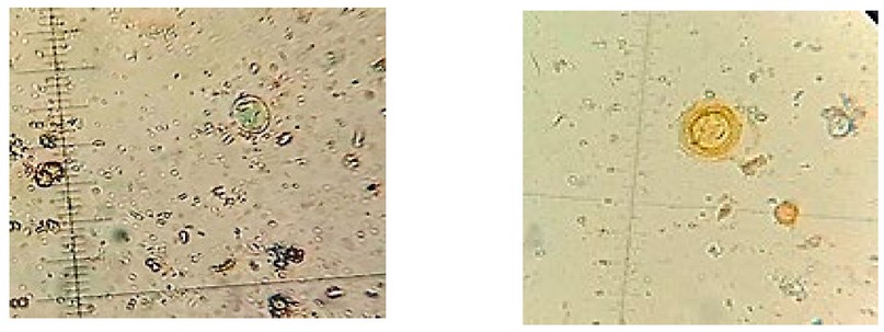
Figure 3. Cyst of Giardia sp Figure 4. Egg of Toxocara vitulorum

Figure 5. Egg of Paramphistomum cervi Figure 6. Egg of Distoma dendriticum
 Figure 7. Egg of Trichostrongylus sp Figure 8. Oocysts of Cyclospora sp
Figure 9 .Cyst of Balantidium coli Figure 10. Cercaria larvae

Figure 11. Filariae
DISCUSSION
Out of the 180 goat fecal samples were collected (one sample for each animal). From the goats examined, 95 (52.77%) were found positive for one or more parasites from Wasit province. The overall higher prevalence of intestinal parasite infection in the areas studied could be attributed to lower host immunity as a result of malnutrition; grazing of young and adult animals together in poorly drained land provide an ideal environment for the transmission of endoparasite eggs to build up a clinical infestation of the host; this finding is consistent with the findings of many other researchers 12 , 13.
According to the data, the prevalence rate of infection with an intestinal parasite is high. It included: Toxocara vitulorum (16.66%), Cryptosporidium spp (11.11%), Amoeba sp (8.8%), Giardia sp (8.8%), Trichostrongylus sp (8.33%), Cyclospora sp (5.55%), Dicrocoelium dendriticum (Distoma dendriticum) (5%), Paramphistomum cervi (4.44%), cercaria larva (2.22%), Balantidium coli (1.66%), Filariae form larvae (1.66%), respectively. This study agreed with 14, who found the highest prevalence of Gastrointestinal parasites in goats (91.55 %) in Kirkuk province, Iraq. And 15 found that infection of goats with gastrointestinal parasites was (81.81 %) in Diyala Province, Iraq. In the current study, the lowest infection rate was observed in December (33.33 %), while the highest was in April (70 %). This study is similar to others 8, 16, 17, which found that the wet months had a higher incidence of infection than the winter months. Maybe a related reason is that reduced grazing hours also reduce the chances of contact between the host and parasites, leading to lower prevalence in the winter season. Further, inclement environmental conditions in winter resulting reduced egg production. According to sex this study recorded infection in females more than in males, agreed with 18, who attributed this variation to a physiological status like pregnancy/ lactation, which causes a dip in natural body resistance in goats against parasites. While 19 and 16 also reported a higher prevalence of gastrointestinal parasites in females than in males. 20 and 21, on the other hand, demonstrated that animal sex did not affect the prevalence of gastrointestinal parasites in small ruminants. In contrast to the current findings, 22 found that males had a higher prevalence and intensity of infection than females. The presence of mixed infection with the gastrointestinal parasites in this study has previously been reported in these animal species 13 , 14, 15, 23. Perhaps the reason is that goats are managed in Conventional systems in which large numbers of animals are routinely kept together. This could be due to increased pasture contamination or to poor sanitation and reduced immunity. This may increase the prevalence of intestinal parasites among the animals.
CONCLUSION
The current study identified several species of intestinal parasites found in goats in Wasit province. This is a significant issue for livestock. As a result, a serious strategy is needed to prevent the spread of more intestinal parasites among local goats And other animals (sheep & cattle) because many parasites may be transmitted between these animals. Furthermore, studies on intestinal parasites in various parts of the country are needed to assess their importance as a source of health hazards. To reduce the parasite burden, some control measures for gastrointestinal parasites in small ruminants must be implemented. Grazing fields should be kept clean and free of animal feces and urine. Education of goat owners on the process of transmission and the influence of these parasites on farm animal productivity should be done regularly.
Acknowledgments
I am grateful to the staff team of Iraq Natural History Research Center and Museum, University of Baghdad, Iraq, for diagnosing the parasites and supplying us the references
Conflicts of Interest
The authors declare no conflict of interest.
REFERENCES
1-Di Cerbo, A.R; Manfredi, M.T; Zanzani, S. ; Stradiotto, K. Gastrointestinal infection in goat farm in Lombardy (Northern Italy): Analysis on community and spatial distribution of parasites. Small Ruminants Research, (2010). 88: 102–112.
2-Dimander, SO; Hoglund, J; Sporndly, E; Waller, P.J. The impact of internal parasites on the productivity of young organically reared on semi-natural pastures in Sweden. Veterinary Parasitology, (2000). 90:271-284. .
3-Johannes, C; Johan, H; Georg, V.S; Pierre, D; Jozef, V. Gastrointestinal nematode infections in adult dairy cattle: Impact on production, diagnosis and control. Veterinary Parasitology, (2009). 164: 70-79.
4-Rahmann, G ; Seip, H. Alternative strategies to prevent and control endoparasite diseases inorganic sheep and goat farming systems–a review of current scientific knowledge. Ressort forschung furden Okologischen Landbau, (2006). 49-90.
5-Jimenez, AE; Montenegro, V.M; Hernandez, J; Dolz, G; Maranda, L; Galindo, J. Dynamicsof infections with gastrointestinal parasites and Dictyocaulus viviparus in dairy and beef cattle from Costa Rica. Veterinary Parasitology, (2007). 148(3-4): 262-271.
5-Jimenez, AE; Montenegro, V.M; Hernandez, J; Dolz, G; Maranda, L; Galindo, J. Dynamicsof infections with gastrointestinal parasites and Dictyocaulus viviparus in dairy and beef cattle from Costa Rica. Veterinary Parasitology, (2007). 148(3-4): 262-271.
6-Kaur, H; Kaur, D. Prevalence of gastrointestinal parasites in domestic animals of Patiala and its adjoining areas. Journal of Veterinary Parasitology, (2008). 22(2): 25-28.
7-Schmidt, G. D; Roberts, L. S; Janovy, J. Foundation of Parasitology. McGrawhill, Boston, Massachusetts,. Science, (2000). pp670.
8-Nwosu, CO; Madu, PP; Richards, W.S. prevalence and seasonal changes in the population of gastrointestinal nematodes of small ruminants in the semi-arid zone of North-Eastern Nigeria. Veterinary Parasitology, (2007) . 144: 118–124.
9-Pedreira, J; Silva, A.P; Andrade, R.S;Suarez, J.L; Arias, M; Lomba, C; Diaz, P; Lopez, C; Banos, P.D ; Morrondo, P. Prevalence of gastrointestinal parasites in sheep and parasite control practices in North-West Spain. Preventive Veterinary Medicine, (2006) . 75: 56-62.
10-Benson, HJ "Microbiological Application: Laboratory Manual in General Microbiology, Short Version",(2002). Eighth Edition, McGraw Hill, Boston MA, USA.
11-Hendrix, C. M; Robinson, E. "Diagnostic Parasitology for Veterinary Technician", (2006). Third Edition, Elsevier Mosby, St. Louis, U.S.A.
12-Asif, M; Azeem, S; Asif, S; Nazir, S. Prevalence of gastrointestinal parasites of sheep and goats in and around Rawalpindi and Islamabad. Pakistan J. Vet. Anim. Sci. (2008) .(1): 14-17.
13-Gadahi, JA; Arshed, M.J; Ali, Q; Javaid, SB; Shah, S.I. Prevalence of gastrointestinal parasites of sheep and goat in and around Rawalpindi and Islamabad. Pakistan Veterinary World, (2009) . 2(2):51-53.
14-Hassan, HF; Barzinji, A.K. Prevalence of Ruminants Gastrointestinal Parasites in Kirkuk province, Iraq, (2018). 13( 3): pp. (96-108)
15-Minnat, TR Detection of gastrointestinal parasite infection of sheep and goats in Diyala Province-Iraq. AL-Qadisiya Journal of Vet. Med. Sci. (2014). 13 (2): 118-123
16-Sharma, DK; Agrawal, N; Mandal, A; Nigam, P; Bhushan, S. Coccidia and gastrointestinal nematode infections in semi-intensively managed Jakhrana goats of semi-arid region of India. Tropical and Subtropical Agroecosystems, (2009). 11: 135- 139.
17-Singh, AK; Das, G; Roy, B; Nath, S; Naresh, R; Kumar, S. Prevalence of gastrointestinal parasitic infections in goat of Madhya Pradesh, India. J. Parasit. Dis., (2015). 39:716-719.
18-Agrawal, N; Sharma, D.K; Mandal, A; Rout, PK; Kushwah, Y.K. Dynamics of faecal egg count in natural infection of Haemonchus Spp. in Indian goats. Vet. World, (2015). 8: 38-41
19-Maqsood, M; Iqbal, Z; Chaudhry, A.H. Prevalence and intensity of haemonchosis with reference to breed, sex and age of sheep and goats. Pak Vet. J., (1996). 16 (1): 41–43.
20-Fikru, R; Teshale, S; Reta, D; Yosef, K. Epidemiology of gastrointestinal parasites of ruminants in Western Oromia, Ethiopia. Int. J. Appl. Res. Vet. M., (2006). 4: 51-57.
21-Tefera, M; Batu, G; Bitew, M. Prevalence of gastrointestinal parasites of sheep and goats in and around Bedelle, South-western Ethiopia. Int. J. Vet. Med., (2009). 8(2): 1-6.
22-Ayaz, MM; Raza, M.A; Murtaza, S; Akhtar, S. Epidemiological survey of helminths of goats in southern Punjab, Pakistan. Trop. Biomed., (2013). 30: 62-71.
23-Bhat, S.A; Rahman Mir, M.U; Qadir, S; Allaie, I.M; Khan, H.M; Husain, I; Sheikh, B.A. Prevalence of gastrointestinal parasitic infections in Sheep of Kashmir valley of India J. Vet. World. (2012) . 5(11):667- 671.
Received: December 23, 2022 / Accepted: January 30, 2023 / Published:15 February 2023
Citation: Makawi Z A Isolation and identification of intestinal parasites from Goats in some areas of Wasit Province, Iraq.Revis Bionatura 2023;8 (1)39. http://dx.doi.org/10.21931/RB/2023.08.01.39