2022.07.04.42
Files > Volume 7 > Vol 7 No 4 2022
Detection of LAP1 and LAP2 genes from Trichophyton rubrum
Hiba Sahib Sadeq1,*, Mouna Akeel Hamed Al-Oebady2
1,2 Biology Department, College of Science, University of Al-Muthanna, Iraq.
* Correspondence author: [email protected] .
Available from: http://dx.doi.org/10.21931/RB/2022.07.04.42
ABSTRACT
Samples of hair, nails and skin were collected, representing people of different ages and races. The number of isolated people gathered between October 2021 and February 2022 from Al-Hussein Teaching Hospital and a private clinic under the supervision of doctors(118) species was examined. Dermatophytes were found in 80 of them; among the 80 positive Trichophyton rubrum species, 30 were produced, which represents less than half of the dermatophytes for each of the 80 positive species (14 cutaneous, nine hair and seven nail isolates)the study's findings included hair hole testing, which came back negative, and urea degradation testing. The results were either negative or inconsistent across the isolates; the growth test in the PDA was positive, the virulence factors that enable the fungus to penetrate host tissues were studied during leucine aminopeptidase (LAP1) and (LAP2), it was observed that there were no significant differences in gene isolates of T. rubrum.
Keywords: LAP1; LAP2; genus Trichophyton rubrum.
INTRODUCTION
Dermatophytes are responsible for various illnesses, including ringworm and ringworm-like infections (Tinea); dermatophytoses is the correct word for a fungus that damages corneal tissues and produces superficial skin diseases such as skin, hair, and nails. It has three principal species: Trichophyton, Microsporum, and Epidermatophyton. Trichophyton spp. is the most frequent Dermatophytoses fungus, with (25) species, the most virulent, and one of the most common diseases in the world, affecting 10- 20% of the global population1.
Many enzymatic components, such as hydrolysis enzymes, are secreted by Dermatophytes, and the higher the ability to create these enzymes, the more ferocity and high ferocity of the attacking fungi, which may cause skin ulcers known as krion, one of the most important enzymes involved in the breakdown of keratin in the host's skin2.
The element mercury is characterized by its high toxicity, especially when inhaled, as it causes scratches and ulcers in the respiratory tract and is mainly produced from industrial processes. Leucine aminopeptidase (LAP1) and (LAP2) are enzymes that help the fungus survive by providing food and preparing the skin for colonization; the presence and production of these enzymes have been investigated genetically in a variety of organisms, including animal cells and some species. Bacteria such as E.coli have been found in the types of genes Trichophyton and Microsporum, but it has not been investigated in depth in sex T.rubrum, even though it is an important marker of the fungus's high virulence3.
MATERIALS AND METHODS
Diagnostic kits
The diagnostic kits used in this work are from the manufacturer (Presto™ Mini gDNA Yeast Kit) and Geneaid/ Germany
Primers
The following primers were used in this work to detect proteins: LAP1(F)TGTCTACAACAACGTCCCCG.and(R)CGTCACCGTCGTAGATC)TGG. The product size is 569 bp, while the LAp2 primer has a length of 487 bp. and the (F)TTGAGTTCCACTGGTACGCC (R) CGACAATGAGCTTAGCGTGC.
Culture media
The following is a list of the various types of media used. The culture media used to isolate and identify fungal isolates were sterilized in an autoclave at 121°C for roughly 15 minutes under 15 psi pressure, and glass was sterilized in an electric oven at 160°C for 90 minutes, per the manufacturer's recommendations2.
Using the SDA (Sabouraud's Dextrose Agar) medium: The dermatophytes isolates were cultured in this medium agar, which was made following the manufacturer's instructions (Liofilchem, Italy) by combining 65 g of media with 1000 ml of distilled water, correcting the pH, and sterilizing everything in an autoclave.
Test medium for urease 1000mL of DW is mixed with the urea agar base. The solution was sterilized in an autoclave, and its pH was set to 7. cooling to 50 °C. A sterile test tube was then filled with the mixture, which had been gently mixed with 50mL of sterilized 40% urea solution. The combination was then allowed to solidify in a slant position. The majority of T. mentagrophytes strains caused the medium to become red from yellow. The color was unaffected by T. rubrum.
Trichophyton Agar No.1 medium was used to differentiate between various fungal isolates. This medium was created following the manufacturer's instructions (Oxoide UK), which included dissolving 59.4 grams in 1000 ml of distilled water, adjusting the pH to 6.8, and sterilizing the medium at 121 °C for 15 min at a pressure of 15.
Medium for Potato Dextrose Agar (PDA) This medium was created following the manufacturer's instructions (Liofilchem, Italy), which called for combining 34 g of PDA with 1000 ml of sterilized water. This medium assessed T. rubrum's capacity to produce a red stain.
Collection of Specimens
A total of (118) samples were collected from patients who visited the medical clinic between October 2021 and February 2022. Skin and its derivatives were used at random in the study, as well as direct diagnosis and transplantation on dishes, using a sterilized sharp blade, samples of skin were scraped from the margin of the infected area, the wounded hair was cut for examination, and the nails were extracted from a sharp blade with pieces of odd shape and color.
Sample transplantation: Samples were transplanted directly into the SDA medium, which contained anti-chloramphenicol and cycloheximide, T. rubrum was identified by planting samples on Sabouraud Dextrose Agar(SDA) and Potato Dextrose Agar (PDA) medium4, 5,6.
Diagnostic tests for Trichophyton rubrum
Hair perforation test: Take baby hair and lay it on a glass slide with a drop of Lacto phenol, then study it under a microscope to see how the fungus attacks the hair if the test is positive7.
Urease test: Depending on the method, this test was used to determine the ability of fungi to make urea enzyme8.
Growing on Trichophyton agar No.1: Trichophyton agar No. 1 is utilized. Agar 1 is a vitamin-free, casein-based control that contains extra histidine. Small, agar-free isolates are moved from SDA plates and cultured at 26oC for 2–3 weeks9,10.
Growth on the medium of the PDA: This medium is used to distinguish T. mentagrophytes from T. rubrum, which produces red dye on the back of the plate. PDA medium is inoculated with the colony at 10-14 days and incubated in the dark at a temperature of 28°C for 2-4 weeks. Rubrum11.
Diagnosis method using PCR assay / by method12.
Hair perforation test: Take baby hair and lay it on a glass slide with a drop of Lacto phenol, then study it under a microscope to see how the fungus attacks the hair if the test is positive 7.
Urease test: Depending on the method, this test was used to determine the ability of fungi to make urea enzyme 8.
Growth on the medium of the PDA: This medium is used to distinguish T. mentagrophytes from T. rubrum, which produces red dye on the back of the plate. PDA medium is inoculated with the colony at 10-14 days and incubated in the dark at a temperature of 28°C for 2-4 weeks. Rubrum 11.
RESULTS
There were 97 positive samples. Fungal infections were found in 80 of the 97 samples. T.rubrum 30 produced positive pieces, accounting for less than half of the cutaneous skin fungus, with 14 isolates from the skin, seven from the nails, and nine from the hair. The following methods were used to identify fungus:
The colony's shape: some colonies were fast-growing, while others were slow-growing; the colony's diameter was around 7 cm in 14 days; the bottom surface seemed red or purple; and many forms of isolates, including fluffy, disintegrated, velvet, and granular isolates. It has a large code production 2.
Micronutrients: Fungi develop little round cones along the hypha in grape clusters, as well as massive conidial with long, smooth, long-stemmed, thin-walled, and helical walls. There is an issue with diagnosing Trichophyton spp genetically, which is called Trichophyton species complex, which is similar to the behavior of species in this species, and the different genotypes may be highly similar in appearance13.
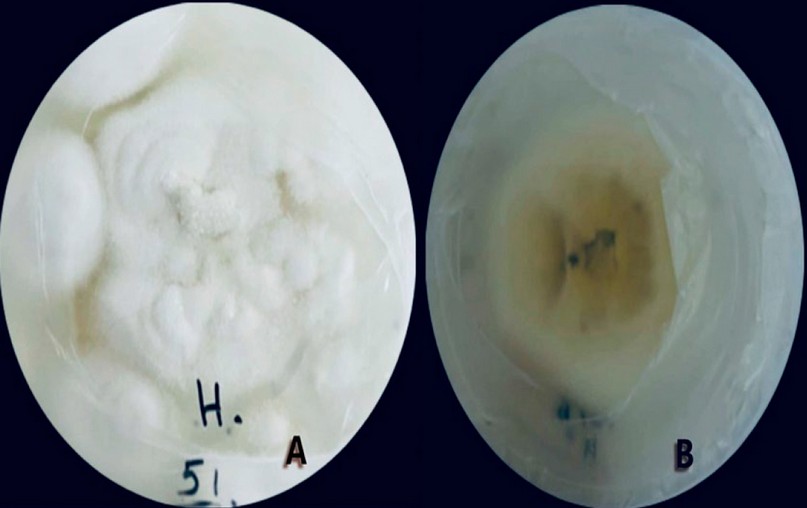
Figure 1. Phenotypic characteristics of T. rubrum and the colony growing on the medium of SDACC, (a) The external appearance from the front (b) The external appearance from the back, temperature 28 ° C, incubation period ten days.
Microscopy revealed a significant number of little conidia that were either sitting or gripping a small bump on the fungal strings in a reciprocal fashion (Teardrop-shaped) or peg-shaped; the huge conidia were quite small and came in a variety of shapes, including pencil-shaped and cigar-shaped conidia, as shown in the following figure (2) , this result agrees with 4,2,14.
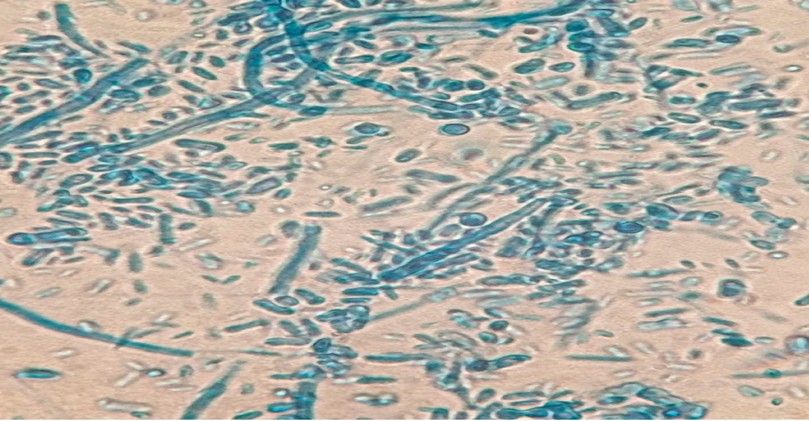
Figure 2. Macroconidia & microconidia of Trichophyton rubrum at 40X with Lacto phenol cotton blue stain.
Physiological tests of the fungus T.rubrum: The hair hole produces a relatively negative result because wall chains developed around the hair blade but not inside it; the urea test showed a negative or mixed effect among the isolates. When growing on the PDA medium, T.rubrum produced a red or pink dye when growing on this medium15.
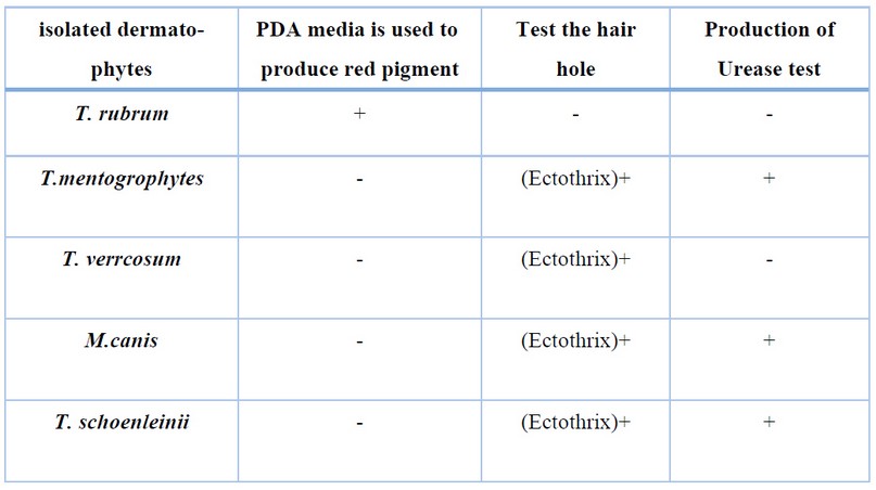
Table 1. Shows the results of fungal physiological tests.
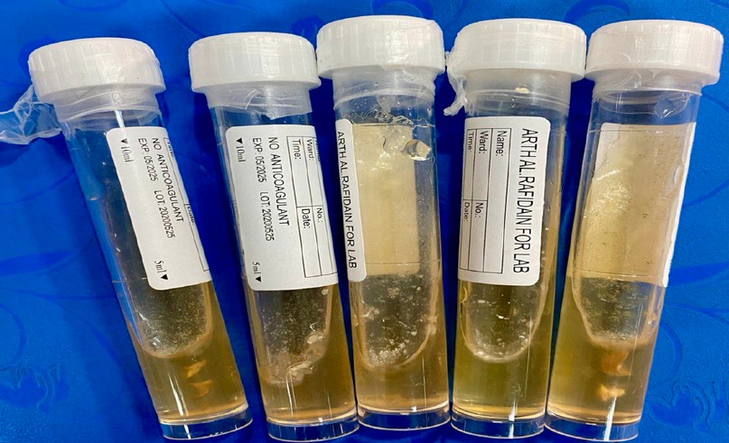
Figure 3. Urea decomposition test. (a) Non-degradation of urea to T. rubrum, the temperature is 28 ℃, and the incubation period is seven days.
The urea test: is a method of determining how much urea when the current findings were compared to those previously reported in the literature, there was a good amount of inconsistency. Negative urease test results were obtained in T. rubrum, whereas positive urease test results were found in the other species; according to the researchers, T. rubrum had positive urease test results, but M. gypseum, M. canis, and T. schoenleinii had negative urease test results8, ,the urease test results for T. mentagrophytes were positive.
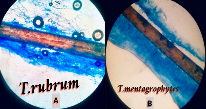
Figure 4. A hair penetration test, (a) No hair penetration in T.rubrum when cultured on SDA medium and at (25 ℃) for an incubation period of (21) days, (b) Hair penetration in T.mentagrophtes when cultured on SDA medium and at (25 ℃) for a period of (21) incubation day
The hair follicle: some dermatophytes produce specialized perforating organs that allow them to penetrate and infect the hair shaft in vitro, while others attack the hair with simple peripheral erosions; as we discovered in this study, the hair perforation test in vitro can distinguish T. rubrum isolates with no perforating organs from atypical T. mentagrophytes separates that produce perforating organs in 8–15 days, and it can also be used to distinguish T. schoenleini 2.
The prevalence of skin fungal infections by place of residence: According to the current study, the incidence of disease with skin fungi was high among patients from rural areas (60 %), whereas the rate of infection in patients from urban areas was low (40 %), as shown in Figure (5), the findings backed up to16, who found that people in rural areas are more affected than city dwellers and that this could be attributed to a lack of hygiene, tinea capitis and tinea corporis have the highest incidence17.
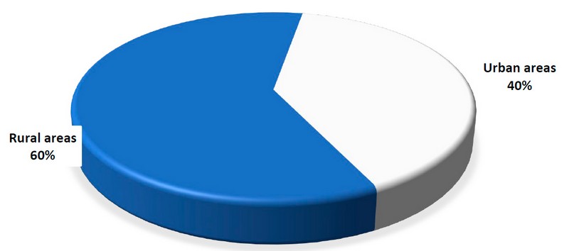
Figure 5. Infection with dermatophytes is distributed according to where the affected people live. *X2 value = 145.664, Significant differences on P≤0.05
Dermatophytes Infection and gender: It has been studied at various ages, from 5 months to 70 years. In the Al-Hussein teaching hospital, it was discovered that males and female dermatophyte infections were converging. Male disorders comprised 61 samples and (51.69%) of the total infection, while female diseases comprised 57 cases and( 48.31%) of the infection. These ratios revealed significant differences in condition between genera, as shown in Figure 6.
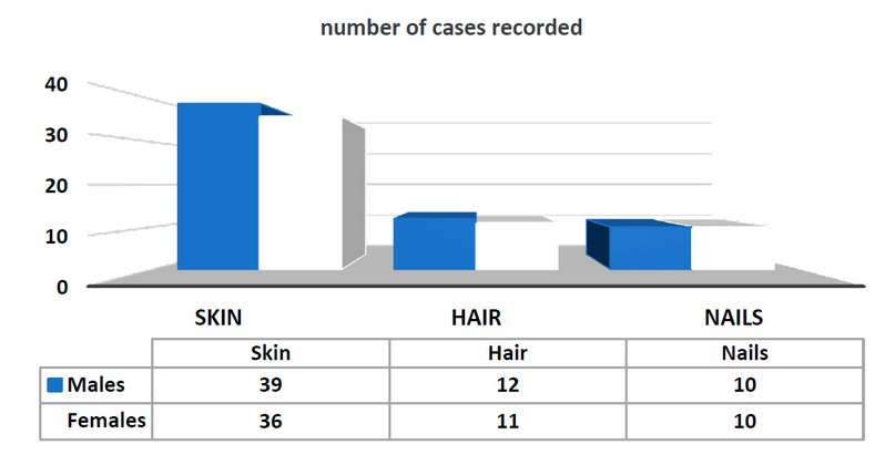
Figure 6. Shows how the number of scales on men's and women's skin, hair, and nails varies. * Not Significant differences on P≤0.05.Error bars represent standard deviation on a significant level of 0.05.
This conclusion was supported by the findings of other researchers, including those from Iraq18.and South Africa19. They discovered that males are more prone to dermatophyte infection than females, in contrast to prior research conducted in water20.
This could be because of the small size of the study sample and the reliance on patients referred to Al-Hussein Teaching Hospital, which had a lot of visitors, or it could be because of the dependence on patients referred to Al-Hussein Teaching Hospital and some clinics, both of which had a lot of visitors. Males are more numerous than females because most girls prefer to visit outpatient clinics.
Dermatophytes Infection associated with Age: The age ranges that were most affected by dermatophytes infection ranged from 5 months to 10 years at (26.66 %), followed by age 11 to 20 years at (18.66 %), age 21 to 30 years at (16.66 %), age 31 to 40 years at (12 %)t, age 41 to 50 years at ( 8.33 %)t, age 51 to 60 years at a ratio of (7.03 %), and age 61 to 70 years at (6.66%).
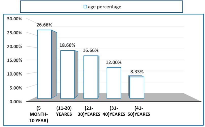
Figure 7. Shows how age and dermatophytes are related. * Significant differences on P≤0.05. Error bars represent standard deviation on a significant level of 0.05.
The findings of this study were consistent with those of numerous previous studies; however, because sample sizes varied, there were a few modest and significant discrepancies at certain times 16.
According to this, dermatophyte infections were more common, and the age group most susceptible to this virus was 5 to 10 years old at a ratio of (80.9 %), which was consistent with the findings of other research. This result was also roughly constant with21. Patients between the ages of 9 and 6 had the highest infection rate. While tinea is more common in children due to a low ratio of fatty acids that prevents fungus growth, it is uncommon in adults due to increased sebaceous gland activity and sebum's antifungal saturated fatty acid content as people age22.
Diagnosis of T.rubrum by Polymerase Chain Reaction (PCR):
The gels were examined under UV light, and one band appeared in all wells at the same level, indicating that primers were bound to their complementary sequence in the DNA template; gel electrophoresis is a technique used to measure the quantity and size of DNA fragments produced after PCR has been completed, the current study's findings are similar to the expected length of about 500 base pairs, the results for the multiplication of a specific primer for dermatophytes T.rubrum, and those of a Korean study23, with a few PCR reaction parameters adjusted that the phenotypic data were similar. The results showed a similarity in the amplified band's molecular weight, which was estimated based on the molecular weights of the band diagnosis was matched by all thirty isolates examined, which is consistent with the findings of24.
T.rubrum was identified molecularly by performing a polymerase chain reaction (PCR) with primers for this (ITS1) and (ITS4), which yielded a 508-base-pair amplification result. This study is not an epidemiological survey but a diagnostic and pathogenic investigation focused on virulence factors, which helped guarantee that the fungus was morphologically accurate. In contrast, isolates from other species were ignored and genetically checked later25.
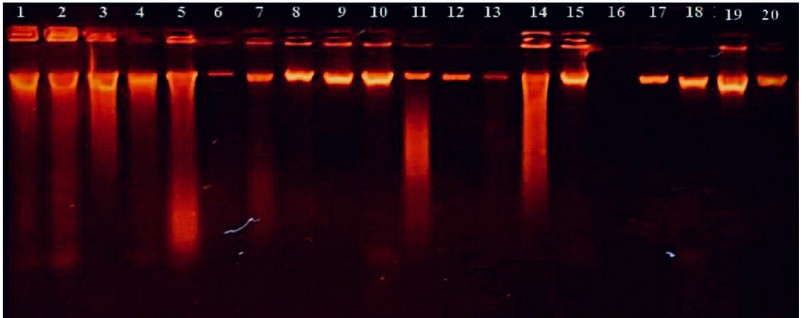
Figure 8. Agarose 1%, 40min. At 110V, stained with Ethidium Bromide agarose gel electrophoresis appearance that displays DNA that was extracted from (human), and for the diagnosis of Trichophyton rubrum, where M: Marker (1500-100bp), represents 1-20 positive fungal isolates for a 508bp assay.
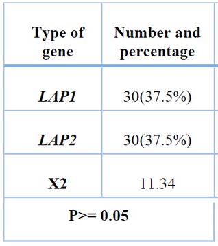
Table 2. Molecular diagnosis of T.rubrum isolates and virulence factors.
Genes Virulence factors
Proteolytic enzymes, which are present in some environmental isolates obtained from soil and organic matter but less frequently, are crucial for the examination of skin components and appendages, especially structural proteins like keratin; their presence is not limited to fungi that attack the stratum corneum of the skin, however; it has been found that some environmental isolates obtained from soil and organic matter contain proteolytic enzymes26, 2.
The temperature and pH of the culture medium, the components of the culture medium, which include sources of carbon, nitrogen, and salts, the duration of incubation, as well as the aeration and fermentation method used, whether it is a surface or submerged fermentation, are just a few of the variables that affect the growth of microorganisms and the production of protease enzymes 2.
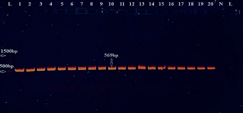
Figure 9. Gel electrophoresis for PCR product of (LAP1 primer) shows 569bp Primer TM at (59), (Agarose 2%, 15min. at 110 volts then lowered to 75 volts for 60min.). They were visualized under UV light after staining with ethidium bromide. Lane L: DNA ladder (100-1500bp).
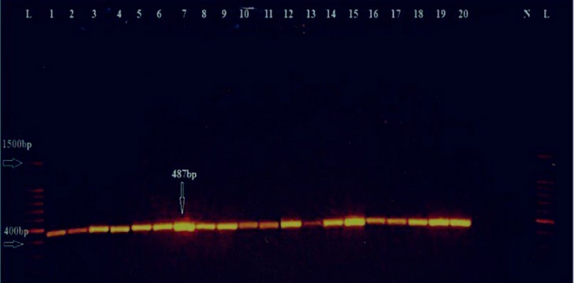
Figure 10. Gel electrophoresis for PCR product of (LAP2 primer) shows 487 bp Primer TM at (57), (Agarose 2%, 15min. at 110 volts then lowered to 75 volts for 60min.). They were visualized under UV light after staining with ethidium bromide. Lane L: DNA ladder (100-1500bp).
DISCUSSION
All ages, particularly children, are affected by fungal infections, which are a major global health issue. As a result, numerous studies on various epidemiological, economic, control, and therapeutic aspects of this infection have been carried out. About 20–25% of the world's population is thought to have skin mycoses, making dermatophytosis one of the most prevalent human fungal illnesses27. The term "dermatophytes" refers to a class of closely related fungi that comprises members of the genera Epidermophyton, Microsporum, and Trichophyton, each of which has several identified species. These fungi are keratinophilic, meaning they attack the skin, hair, and nails of humans and animals28.
Most dermatophyte strains previously had a relatively narrow geographic distribution, making them one of the few fungi that cause infectious diseases. On the other hand, dermatophytosis has recently become one of the most prevalent infectious disorders affecting people worldwide and is spread globally. As many other disorders resemble the clinical presentation, it is challenging to identify dermatophytosis based solely on clinical signs. Seborrheic dermatitis, atopic dermatitis, contact dermatitis, psoriasis, candidal intertrigo, erythrasma, eczema, etc., are among the dermatophytoses that can be diagnosed as a differential diagnosis29.
Additionally, immunocompromised patients, such as those with AIDS, diabetes mellitus, organ transplantation, corticosteroid use, and antineoplastic agent use, are more challenging to diagnose for dermatophytosis.In order to apply the proper treatment and preventative measures, reliable laboratory methods must be available for the quick and accurate identification of the dermatophytes involved. The conventional approaches to finding fungi have limitations, such as the low specificity of KOH microscopy and the low sensitivity and lengthy nature of fungus culture. The results of culture isolation are further compromised because additional dermatophyte isolates from patients receiving antifungal treatment typically do not exhibit characteristic morphology on culture. This study's few negative results from direct microscopic examination and culture could be related to several factors, including the small amount of collected sample, which might not have been enough to produce a positive result. Because many people with dermatophytoses utilized random drugs without seeing a doctor, the adverse effects of culturing specimens may be explained. Instead of causing them to expand, this led to a shift in the dermatophytes' vitality, perhaps due to improper sample storage before culture. Storage in containers that retain moisture causes saprophytic fungus to develop, contaminating the original sample and yielding unfavorable results. The hyphae of non-dermatophytic fungi, such as molds, which frequently only appear as temporary contaminants and are not the trustworthy causative agent of the disease, are very difficult to distinguish from those of dermatophyte hyphae30.
Tinea cruris was the third most common kind of dermatophytosis in our study, Adults in the (31-50) year age range were impacted, and more men than women experienced it. According to Rothman, 1985, ringworm of the groin is primarily a postpubertal disease of men and is most likely caused by the pubertal development of a sex-specific apocrine gland, whose secretion contributes to infection susceptibility. According to data, urban areas had a 40% infection rate, while rural areas had a 60% infection rate. However, given the predominance of persons who live in urban settings, their findings differ from those of others. This can be explained by the fact that many rural places have poor health, financial difficulties, and congestion of residents 31.
The initial PCR technique is DNA fingerprinting. It has been demonstrated that this primer is an effective tool for the molecular identification of dermatophytes. In this study, we successfully classified T.rubrum isolates as dermatophyte species32.
CONCLUSIONS
The most common species that induced dermatophytoses were Trichophyton rubrum, Trichophyton mentagrophytes, and Microsporum canis. The T.rubrun fungus outnumbers other species that cause skin infections, indicating that it is very pathogenic. Anthropophilic disorders were more prevalent than other categories, and animal interaction and poor hygiene in rural areas were the main causes of dermatophytes infection; the fungus T. rubrum carries the genes LAP and LAP2, which produce proteolytic enzymes that aid in adhesion, invasion, and suppression of the host response, making the fungus more likely to colonize and penetrate tissues.
Funding: Self-funding.
Conflicts of Interest: There is no conflict.
REFERENCES
1. Brescini L, Fioriti S, Morroni G, Barchiesi F. Antifungal combinations in dermatophytes. J Fungi. 2021;7(9). doi:10.3390/jof7090727
2. Al-Shibly, M. K., & Al-Marshedy, A. M. Molecular study of virulence factors influencing the pathogenicity of Trichophyton rubrum. Eco. Env. & Cons.2019., 25(1), 153-158..
3. Matsuietal, BiolChem (2).2006.
4. Tambor JHM, Guedes RF, Nobrega MP, Nobrega FG. The complete DNA sequence of the mitochondrial genome of the dermatophyte fungus Epidermophyton floccosum. Curr Genet. 2006;49(5):302-308. doi:10.1007/s00294-006-0057-2
5. Xu Y, Hall C, Wolf-Hall C, Manthey F. Fungistatic activity of flaxseed in potato dextrose agar and a fresh noodle system. Int J Food Microbiol. 2008;121(3):262-267. doi:10.1016/j.ijfoodmicro.2007.11.005
6. Kannan, P., Janaki, C., & Selvi, G. S.. Prevalence of dermatophytes and other fungal agents isolated from clinical samples. Indian Journal of Medical Microbiology.2006, 24(3), 212-215..
7. Rey, L. Revista do Instituto de Medicina Tropical de São Paulo (Journal of the São Paulo Institute of Tropical Medicine) fifty years later. Revista do Instituto de Medicina Tropical de São Paulo.2009, 51, 239-240..
8. Padhye, A. A., & Ajello, L. The taxonomic status of the hedgehog fungus Trichophyton erinacei. Sabouraudia.1977, 15(2), 103-114..
9. Geber A, Hitchcock CA, Swartz JE, et al. Deletion of the Candida glabrata ERG3 and ERG11 genes: Effect on cell viability, cell growth, sterol composition, and antifungal susceptibility. Antimicrob Agents Chemother. 1995;39(12):2708-2717. doi:10.1128/AAC.39.12.2708
10. Abarca, M. L., Castellá, G., Martorell, J., & Cabañes, F. J. Trichophyton erinacei in pet hedgehogs in Spain: Occurrence and revision of its taxonomic status. Medical Mycology.2017, 55(2), 164-172..
11. Akram, M., Shahid, M., & Khan, A. U. Etiology and antibiotic resistance patterns of community-acquired urinary tract infections in JNMC Hospital Aligarh, India. Annals of clinical microbiology and antimicrobials.2007, 6(1), 1-7..
12. Jousson, O., Lechenne, B., Bontems, O., Capoccia, S., Mignon, B., Barblan, J., ... & Monod, M. 2004.
13. S. M. Abdulateef, O. K. Atalla1, M. Q. A L-Ani, TH. T Mohammed, F M Abdulateef And O. M. Abdulmajeed. Impact of the electric shock on the embryonic development and physiological traits in chicks embryo. Indian Journal of Animal Sciences.2021, 90 (11): 1541–1545.
14. Gibas, C. F. C., Sigler, L., Summerbell, R. C., Hofstader, S. L. R., & Gupta, A. K.. Arachnomyces kanei (anamorph Onychocola kanei) sp. nov., from human nails. Medical Mycology.2002, 40(6), 573-580..
15. Aala F. Conventional and molecular characterization of Trichophyton rubrum. African J Microbiol Res. 2012;6(36):6502-6516. doi:10.5897/ajmr10.736
16. Fathy H, El-Mongy S, Baker NI, Abdel-Azim Z, El-Gilany A. Prevalence of skin diseases among students with disabilities in Mansoura, Egypt. East Mediterr Heal J. 2004;10(3):416-424. doi:10.26719/2004.10.3.416
17. Bongomin F, Gago S, Oladele RO, Denning DW. Global and multi-national prevalence of fungal diseases—estimate precision. J Fungi. 2017;3(4). doi:10.3390/jof3040057
18. Ali, T. H., Ali, N. H., & Mohamed, L. A. Production, Purification And Some Properties Of Extracellular Keratinase From Feathers-Degradation By Aspergillus Oryzae Nrrl-447. Journal of Applied Sciences in Environmental Sanitation.2011, 6(2)..
19. Islam, T. A. B., Majid, F., Ahmed, M., Afrin, S., Jhumky, T., & Ferdouse, F. Prevalence of dermatophytic infection and detection of dermatophytes by microscopic and culture methods. Journal of Enam Medical College.2018, 8(1), 11-15..
20. Kobylak N, Bykowska B, Nowicki R, Brillowska-Dabrowska A. Real-time PCR approach in dermatophyte detection and Trichophyton rubrum identification. Acta Biochim Pol. 2015;62(1):119-122. doi:10.18388/abp.2014_864
21. Mohammed, S. J., Noaimi, A. A., Sharquie, K. E., Karhoot, J. M., Jebur, M. S., Abood, J. R., & Al-Hamadani, A.. A survey of dermatophytes isolated from Iraqi patients in Baghdad City. Al-Qadisiyah Medical Journal.2015, 11(19), 10-15..
22. Mohammed, S. J. A Survey of Dermatophytes Isolated from Cows and Sheep in Iraq: Sudad Jasim Mohammed, Mohammed K Faraj. The Iraqi journal of veterinary medicine.2011, 35(2), 40-45..
23. Warrilow, A. G., Parker, J. E., Price, C. L., Garvey, E. P., Hoekstra, W. J., Schotzinger, R. J., ... & Kelly, S. L. The tetrazole VT-1161 is a potent inhibitor of Trichophyton rubrum through its inhibition of T. rubrum CYP51.2017.
24. Othman Ghazi Najeeb Alani , Yassen Taha Abdul-Rahaman and Thafer Thabit Mohammed. Effect Of Vêo® Premium and Vitamin C Supplementation on Lipid Profile Before and During Pregnancy in Some Local Iraqi Ewes During Heat Stress. Iraqi Journal of Science.2021, Vol. 62, No. 7, pp: 2122-2130.
25. De Pauw, B., Walsh, T. J., Donnelly, J. P., Stevens, D. A., Edwards, J. E., Calandra, T., ... & Bennett, J. E.2008.
26. Turpie AG, Lassen MR, Davidson BL, et al. Rivaroxaban versus enoxaparin for thromboprophylaxis after total knee arthroplasty (RECORD4): a randomised trial. Lancet. 2009;373(9676):1673-1680. doi:10.1016/S0140-6736(09)60734-0
27. Havlickova B, Czaika, ViktHavlickova B, Czaika VA, Friedrich M. Epidemiological trends in skin mycoses worldwide .2008, 51, SUPPL. 4, (2-15)). Mycoses. 2009;52(1):95. doi:10.1111/j.1439-0507.2008.01668.x
28. Eman-abdeen, W. S. M. overview on bovine dermatophytosis.2018.
29. Barnes, P. J. Theophylline: new perspectives for an old drug. American journal of respiratory and critical care medicine.2003, 167(6), 813-818..
30. Dasgupta, S., Das, S., Chawan, N. S., & Hazra, A.. Published online 2020:1-4.
31. Abu-Elteen, K. H., & Abdul Malek, M. Prevalence of dermatophytoses in the Zarqa district of Jordan. Mycopathologia.1999, 145(3), 137-142..
32. Faggi, E., Pini, G., Campisi, E., Bertellini, C., Difonzo, E., & Mancianti, F. Application of PCR to distinguish common species of dermatophytes. Journal of Clinical Microbiology.2001, 39(9), 3382-3385..
Received: August 25, 2022 / Accepted: October 12, 2022 / Published:15 November 2022
Citation: Sadeq H S, Al-Oebady M A H. Detection of LAP1 and LAP2 genes from Trichophyton rubrum. Revis Bionat a 2022;7(4) 42. http://dx.doi.org/10.21931/RB/2022.07.04.42
