2023.08.01.85
Files > Volume 8 > Vol 8 No 1 2023
Measuring levels of hormones in osteoporosis Iraqi women patients

Ali Kareem Juhi 1, Aliaa Saadoon Abdul- Razzaq Al-Faraji 2,*
1 Affiliation: Department of Chemistry / Collage of Sciences /University of Baghdad/ Baghdad/Iraq.
2 Affiliation: Market Research and Consumer Protection Center/University of Baghdad/ Baghdad/Iraq. [email protected]
* Corresponding author: [email protected]
Available from: http://dx.doi.org/10.21931/RB/2023.08.01.85
ABSTRACT
This study was conducted to investigate the effect of the hormone progesterone and testosterone on osteoporosis in women, and it was found that there are statistically significant differences between the hormone progesterone and there are no statistically significant differences for the hormone testosterone. The ELISA method measured the testosterone concentration in the blood and the progesterone concentration. The purpose of the study measuring the levels of some hormones in osteoporosis Iraqi women patients. Fifty (50) women patients with osteoporosis were admitting Baghdad Teaching Hospital with osteoporosis and 40 healthy populations, and it was conducted in Baghdad Teaching Hospital/ Bone density examination unit/ in Baghdad-Iraq. Endocrine testing Estrogen content was measured following the manufacturer's instructions using available human Uno ELISA kits (Immunolab GmbH, Kassel, Germany). Results showed a significant difference (P-value of 0.0038) in progesterone between the two study groups. The mean of patient groups was mean±SD (6.759 ± 6.705), and control groups were mean±SD (11.03 ± 6.546). The main risk factor for osteoporosis is the presence of more progesterone when comparing patients with healthy women. As for testosterone, there are no significant differences; that is, it does not affect women
Keywords: ELISA Technique, progesterone, testosterone
INTRODUCTION
Primary osteoporosis is caused by loss of evil and a disturbance of bone architecture, resulting in skeletal weakness and increasing fracturing risk 1. Vertebral, hip, and other fragility fractures in women, which are linked to fractures in old age, originate in the pre-and peri-menopausal age, when a range of variables influencing bone mineral metabolism are present. 2 .anti characteristics such as female species, elderly aging, tiny thin build, ethnicity, and a family history of cracks are among them. Vitamin d. insufficiency, inactivity, alcoholism, excess drinking, & caffeine are all key variable drivers 3. Quasi and new therapies are used to protect and cure osteo. Diet, regular weight-bearing activities, and the cessation of smoking and alcohol intake are five semi-therapeutic factors that have been well documented. The World Health Organization's (WHO) concept of fractures is as follows:4. The criteria provide the clinician with an accurate benchmark to definitively diagnose and make subsequent treatment strategies for the younger typical sample population (table 1) 5. Moreover, the disease is known as a "motionless disease," so it progresses commences; causes severe pain, lower quality of life, lost workdays, and disability; and approximately 20% of women who have a hip fracture will drop dead within a week as an indirect result of both the fracture.
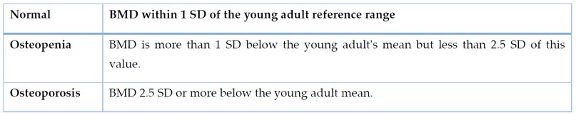
Table 1. The definition of osteo by the Health Organisation (WHO).
Testosterone is one of a man's major hormones. We also know that this hormone level tends to decline. The necessity of monitoring testosterone levels in adult males is a lesser-known truth. They will indeed be able to intervene in the aging process in this way. The above are a few truths about testosterone and how to replace it if it is low. I'll also talk about how the medical profession deals with this. Some people are scared of hrt 6 .steroid.
On the other hand, drugs and estrogen act in tandem in every female tissue. Fractures decrease when testosterone production falls. Inflammatory mediators produce postpartum reduced BMD, which leads to more significant demineralization in women 7.
MATERIAL AND METHODS
Patients Group
Fifty (50) women patients with osteoporosis were admitting Baghdad Teaching Hospital with osteoporosis and 40 healthy populations and it was conducted in Baghdad Teaching Hospital/ Bone density examination unit/ in Baghdad-Iraq in the period between September 2020- January 2021
Control Group
It consisted of Forty (40) healthy volunteers. All patients and control groups were from the same ethnic group (Arabic).
(ELISA): Enzyme-Linked Immunosorbent Assay (Analysis)
Endocrine testing Estrogen content was measured following the manufacturer's instructions using available human Uno ELISA kits (Immunolab GmbH, Kassel, Germany). A calibration curve of 17-estradiol, estrogen, and hormone (area" levels) was injected in the microwells as we prepared solutions of blood serum. The dissolved enzyme conjugate (peroxidase), substrate (tetramethylbenzidine -TMB), and antibodies were added to the microplate and incubated in the dark for 30 min. Serological washing was used to wash all precedes in stages (ELx50, Bio-Tek Instruments, Germany). The solution was allowed to cool to a stop solution (0.5 M sulfate). The serum hormone concentration was determined by drawing the calibration graph. 8
RESULT
Distribution of Osteoporosis Patients and Control Group According to progesterone
Results of osteoporosis patients (women) and an apparently healthy group were studied according to the progesterone table 2 and figure 2. The results showed a significant difference (P-value of 0.0038) in progesterone between the two study groups. The mean of patient groups was mean±SD (6.759 ± 6.705), and control groups were mean±SD (11.03 ± 6.546). 9,10

Figure 1. Distribution of Osteoporosis Patients and Control Groups According to progesterone.

Table 2. Distribution of Osteoporosis Patients and Control Groups According to Testosterone.

DISCUSSION
Results of osteoporosis patients (women), as well as a healthy group, were studied according to the Testosterone table (2) and figure (1). The results showed a non-significant difference (P-value of 0.2785) in testosterone between the two study groups. The mean of patient groups was mean±SD (12.30± 3.677), and control groups were mean±SD (11.40± 4.165) 11,14,15
CONCLUSION
The main risk factor for osteoporosis is the presence of more progesterone when comparing patients with healthy women. As for testosterone, there are no significant differences; it does not affect women.
Ethics Approval and Consent to Participate
According to human research's local bioethical principles, anonymized, unidentifiable data from clinical records, excluding case reports, do not require internal review boards' approval. The physicians collecting clinical data were the only health providers accessing patients' clinical records.
Competing Interests
The authors declare no conflicts of interest.
Funding
This study did not receive any funding.
Authors' Contributions
ASR conceptualized the study and directed the team when collecting information. He drafted the first version of the manuscript and reviewed the final version. ASR and AKJ collected information from the Iraqi woman unit and contributed equally to the data analysis. ASR was responsible for critically reviewing the first draft, completing the manuscript's final version, and critically reviewing the entire analytical process around data collection.
REFERENCE
1 Lane, N.E. Epidemiology, etiology, and diagnosis of osteoporosis. American journal of obstetrics and gynecology, 2006. 194(2): p. S3-S11.
2 Suwaid, A. H. .; Rashid, M. A. .; Taha, M. M. . Genetic Analysis For Combining Ability And Estimation Of Some Genetic Parameters Of Yield And Its Components In Maize Using Half Diallel Cross. ). Journal of Life Science and Applied Research. 2020, 1, 60-64..
3 Malhotra, N.; Mithal, A.;Dewq, R.Q. Osteoporosis in Indians. Indian Journal of medical research, 2008. 127(3).
4 Lewiecki E., Clinical applications of bone density testing for osteoporosis. Minerva medica, 2005. 96(5): p. 317-330.
5 Jang H.-D.Relationship between bone mineral density and alcohol intake: A nationwide health survey analysis of postmenopausal women. PLoS One, 2017. 12(6): p. e0180132.
6 Wulandari P.The Profile of Progesterone Hormone, Vitamin D, and Bone Density in Postmenopausal Women. Journal of International Dental and Medical Research, 2019. 12(3): p. 842-847.
7 Pal R., Agrawal K., Gupta S. et al .worsening of unrecognized tumour-induced osteomalacia with inadvertent use of recombinant human parathyroid hormone. Endokrynol Pol,2020; 71:102–103. https:// doi. org/ 10. 5603/ EP. a2019. 0045
8 Morris, H.A.; Eastell, R.; Jorgensen, N.R;.Clinical usefulness of bone turnover marker concentrations in osteoporosis. Clin Chim Acta,2017. 467:34–41. https:// doi. org/ 10. 1016/j. cca. 2016. 06. 036.
9 Díez-Pérez, A.; Marin, F.;Eriksen, E.F. Effects of teriparatide on hip and upper limb fractures in patients with osteoporosis: a systematic review and meta-analysis. Bone ,2019.120:1–8. https:// doi. org/ 10. 1016/j. bone. 2018. 09. 020
10 Kanis, J.A.; Johansson, H.;Odén, A.;Realm, T.S.Characteristics of recurrent fractures. Osteoporos Int .2018.29:1747–1757. https:// doi.org/ 10. 1007/ s00198- 018- 4502-0
11 Bansal, B.; Mithal, A.; Chopra, S.R. Judicious use of DXA-BMD in assessing fracture risk by using clinical risk factors in the Indian population. Arch Osteoporos .2018.13. https:// doi.org/ 10. 1007/ s11657- 018- 0536-3.
12 Garg, A.; Aggarwal, A.;Pal, R.;Ser, AX eTrabecular bone score in healthy adult population of India: Chandigarh Urban Bone Epidemiological Study (CUBES). J Endocr Soc 3..2019. https:// doi. org/ 10. 1210/ js. 2019- SAT- 53773.
13 Sooragonda, B.; Cherian, K.E.; Jebasingh, F.K.;, Che,C.Z.Longitudinal changes in bone mineral density and trabecular bone score following yearly zoledronic acid infusion in postmenopausal osteoporosis—a retrospective-prospective study from southern India. Arch Osteoporos,2019. 14. https:// doi. org/ 10. 1007/s11657- 019- 0630-1
14 Leder, B.Z. Optimizing sequential and combined anabolic and antiresorptive osteoporosis therapy: optimizing anabolic and antiresorptive therapy. JBMR Plus 2018.2:62–68. https:// doi. org/ 10.1002/ jbm4. 10041
15 Alkubaisy,S.A., A.A. Majid, S.M. Abdulateef, F.A. Al-Bazy, O.K. Attallah, O.M. Abdualmajeed, Th. T. Mohammed, F.M. Abdulateef, K.I. Mahmud. Effects of In-Ovo injection of Biotin on chick's embryonic development and physiological traits. IOP Conference Series: Earth and Environmental Science.2021, 761(1), 012111.
Received: January 15, 2023 / Accepted: February 25, 2023 / Published:15 March 2023
Citation: Juhi, AK; Al-Faraji, A.S.A.; Measuring levels of hormones in osteoporosis Iraqi women patients.Revis Bionatura 2023;8 (1) 85. http://dx.doi.org/10.21931/RB/2023.08.01.85