2024..01.01.58
Files > Conference Series > 2024 > Chimboazo ild pagina nueva
Ameliorative Effect of Cinnamon and Rosemary Oils in Acrylamide–Induced Hepatic Injury in Rats
Hala Elsayed1*, Ashraf Abd El-Hakim El komy2, Elham Abd-El Moneim El-Shewy3, Faten Ebrahim Elsayed Abdallah4
1Department of Pharmacology, Faculty of Veterinary Medicine, Benha University, Toukh 13736, Egypt.; [email protected].
2Department of Pharmacology, Faculty of Veterinary Medicine, Benha University, Toukh 13736, Egypt.; [email protected].
3Department of Forensic Medicine Faculty of Veterinary Medicine, Benha University, Toukh 13736, Egypt.; [email protected].
4Department of Pharmacology, Faculty of Veterinary Medicine, Benha University, Toukh 13736, Egypt.; [email protected].
* Correspondence: [email protected].; Tel.: +20 102 608 7482.
Available from. http://dx.doi.org/10.21931/BJ/2024.01.01.58
ABSTRACT
Liver diseases can result from various causes, such as viruses, bacteria, autoimmune disorders, or certain medications and toxic substances. While modern medicine offers treatments for these conditions, there needs to be more effective drugs that can protect and regenerate liver cells. Therefore, it is crucial to identify new treatment options and liver-protective agents that are both highly efficient and safe. This study is assigned to investigate the adverse effects of acrylamide on the liver in rats and explore whether these effects can be mitigated by co-administration of cinnamon oil (C.O.), rosemary oil (R.O.), or a combination of both oils during acrylamide exposure. A total of 70 male albino rats were divided randomly into 7 groups, each group of 10 rats, that received different treatments: control group, acrylamide-treated group (20 mg/kg b.wt), cinnamon oil-treated group (200 mg/kg b.wt), rosemary oil-treated group (250 mg/kg b.wt), acrylamide and cinnamon oil-treated group, acrylamide and rosemary oil-treated group, and acrylamide, cinnamon oil, and Rosemary oil-treated group. These treatments were administered orally for 28 consecutive days. Blood and liver tissue samples were gathered at the end of the study to assess the outcomes. The results revealed that cinnamon oil and rosemary oils exhibited hepatoprotective effects, as evidenced by normalized liver function parameters (alanine transaminase, Aspartate transaminase, and Alkaline phosphatase), as well as improvements in non-enzymatic parameters (total protein, albumin, cholesterol, triglyceride, low-density lipoprotein, and high-density lipoprotein). The observed hepatoprotection of cinnamon oil and rosemary oils was attributed to their ability to reduce oxidative stress caused by acrylamide, as demonstrated by lower levels of liver cell lipid peroxidation product (malondialdehyde) and enhanced activity of antioxidative enzymes (glutathione and catalase) in liver tissue.
Keywords: Cinnamon, Rosemary, Acrylamide, Liver, Rats, Antioxidants.
INTRODUCTION
Acrylamide is a compound that dissolves in water and is extensively utilized in various fields, including dye production, soil coagulation, wastewater treatment, paper packaging, and laboratory applications 1. This compound is not naturally present. Research has revealed that foods with high carbohydrate content, when exposed to high temperatures during heating or frying processes, tend to contain significant levels of acrylamide 2. Acrylamide forms when carbohydrate-rich foods like crackers, potatoes, crisps, cereals, bread, and French fries are grilled, fried, baked, or roasted at temperatures exceeding 120°C. This process involves the reaction between the amino acid asparagine and a reducing sugar such as glucose 3.
Acrylamide has been associated with several harmful effects in rodents, including genotoxicity, neurotoxicity, hepatotoxicity, nephrotoxicity, carcinogenicity, and reproductive toxicity 4. The International Agency for Research on Cancer has classified acrylamide as a 2A substance 5. From the Lauraceae family, Cinnamon is a widely used spice in the food industry, often added to baked goods and chili sauce for flavoring purposes 6. It contains various constituents that act as food additives, providing antioxidant, anti-inflammatory, anti-cancer, antimicrobial, and antidiabetic properties 7. From the Lamiaceae family, Rosemary is a well-known aromatic plant used for flavoring in food processing and medicinal purposes 8. Rosemary is recognized for its potent antioxidant properties, which help eliminate free radicals, strengthen the antioxidant system, and prevent oxidative stress 9.
Rosemary extracts have been reported to possess hepatoprotective activity in a rat model of azathioprine-induced toxicity and acetaminophen-induced liver damage. When administered to rats with hepatotoxicity, Cinnamon and Rosemary or their combination resulted in a notable reduction in the average level of MDA and a significant increase in hepatic GSH.9 Currently, the toxic effects of acrylamide on liver tissue in animals have been examined, along with the evaluation of the beneficial effects of Cinnamon and Rosemary in relieving acrylamide-induced hepatotoxicity in adult albino rats.
MATERIALS AND METHODS
Acrylamide, a white crystalline powder obtained from Fine–Chem Limited Company with a purity of approximately 98.5%, was dissolved in distilled water just before administration at a dose of 20 mg/kg body weight 10.
Cinnamon and rosemary oils, sourced from El-captain Company for extracting natural oils, herbs, and cosmetics in Cairo, Egypt, were used at 200 mg/kg body weight 11 and 250 mg/kg body weight 12, respectively.
Various diagnostic kits were used for measuring levels of aspartate aminotransferase (AST), alanine aminotransferase (ALT), alkaline phosphatase (ALP), total protein, albumin, triglycerides, cholesterol, HDL, LDL, as well as oxidative stress markers including malondialdehyde (MDA), glutathione peroxidase (GSH), and catalase (CAT) in plasma and liver tissue homogenate. These kits were obtained from BioDiagnostic Company in Giza, Egypt.
The study utilized 70 male white albino Wistar rats of about 180-200 grams. These rats were brought from the Center of Laboratory Animals at the Faculty of Veterinary Medicine, Benha University, Egypt. Before the experiment, the rats were given one week to acclimatize. During this period, they were provided with a standard laboratory-balanced commercial diet and had access to water ad libitum. The research adhered to the guidelines for animal care and procedures set by the Faculty of Veterinary Medicine, Benha University, Benha, Egypt, under number (03-03-23). The basal diet provided to the rats met their nutritional requirements for maintenance as specified by the National Research Council and Reeves.
The acclimatized rats were divided into seven groups, each consisting of 10 rats. The groups were treated differently according to the objective of study 10. The treatments were as follows:
· Group 1: Control group, where rats were given distilled water orally once a day for 28 consecutive days.
· Group 2: Rats given acrylamide orally at a dose of 20 mg/kg body weight once daily for 28 consecutive days to induce hepatic injury 9.
· Group 3: Rats given cinnamon oil orally at 200 mg/kg body weight once daily for 28 consecutive days 10.
· Group 4: Rats given rosemary oil orally at a dose of 250 mg/kg body weight once a day for 28 consecutive days 11.
· Group 5: Rats were given acrylamide (20 mg/kg body weight) and cinnamon oil (200 mg/kg) daily for 28 consecutive days.
· Group 6: Rats were given acrylamide (20 mg/kg body weight) and rosemary oil (250 mg/kg) daily for 28 consecutive days.
· Group 7: Rats that were given acrylamide (20 mg/kg body weight) followed by cinnamon oil (200 mg/kg body weight) and then rosemary oil (250 mg/kg body weight) once a day for 28 consecutive days.
Sampling
At the end of the 28-day treatment period, all rats were euthanized. Blood and liver samples were collected to conduct biochemical and histopathological examinations.
Blood samples were obtained from 10 rats in each group after 28 days of treatment by puncturing the retro-orbital plexus, which is located at the inner corner of the eye. Capillary tubes were used to collect the blood samples and immediately placed in plain sampling tubes. The samples were then centrifuged at 1200 g for 10 minutes to separate the clear plasma, which was transferred into Eppendorf tubes using automatic pipettes. The plasma samples were stored at -20 °C for further analysis.
Tissue specimens
The liver underwent an examination and rinsing process using a standard saline solution to remove traces of blood, fat, and connective tissue. Liver samples of approximately 1 gram were carefully collected from each group of 10 rats and placed into secure and labeled Eppendorf tubes. These samples were blended with a pH 7.4 phosphate buffer solution and then centrifuged at a speed of 1200 g for 10 minutes. The resulting clear liquid was carefully preserved at -20°C for further analysis of the oxidative markers in the tissues. Moreover, additional liver tissue samples were obtained and maintained in a 10% formalin solution for histopathological examinations following a 28-day treatment period.
Biochemical and antioxidant effects
The levels of certain enzymes in blood and liver tissue homogenates were determined to evaluate the drug's safety. The plasma activities of AST, ALT, and ALP were measured using specialized kits and the methods outlined by Reitman & Frankel 12 for ALT and ALT and by Haussament 13 for ALP, following the instructions provided by the manufacturer. Other biochemical parameters such as total protein, albumin, T.G.s, HDL cholesterol, and LDL were also measured using specific spectrophotometric methods 13. Furthermore, the oxidative and antioxidative parameters in liver homogenate were determined using particular kits for measuring MDA, GSH, and Catalase through spectrophotometry 14.
Histopathological examination was conducted according to Bancroft 15.
Data management and statistical analysis
The collected data were analyzed using one-way analysis of variance (ANOVA), and post hoc testing was performed using Duncan's test. The study was conducted using the Windows version of SPSS 25. The results were presented as mean±standard error (SEM), and statistical significance was considered at P values ≤0.05.
RESULTS
The results of the biochemical parameters analysis revealed that rats treated with acrylamide exhibited a significant rise in AST, ALT, ALP, cholesterol, triglycerides, and LDL-cholesterol levels and a significant decrease in total protein, albumin, and HDL cholesterol levels compared to the control groups. However, the administration of Cinnamon and Rosemary led to a notable improvement in these parameters, as depicted in Tables 1 and 2. Regarding oxidative stress results (Figures 1, 2, and 3), acrylamide-treated rats demonstrated a marked increase in MDA levels and a significant reduction in CAT and GSH levels in their liver tissues compared to the control groups. However, the administration of Cinnamon and Rosemary resulted in a considerable improvement in these parameters. Furthermore, a histopathological examination of the liver tissues from the acrylamide-treated group showed changes in their structural appearance. Nevertheless, an enhancement in histopathology was observed after the administration of Cinnamon and Rosemary, as shown in Figure 4.
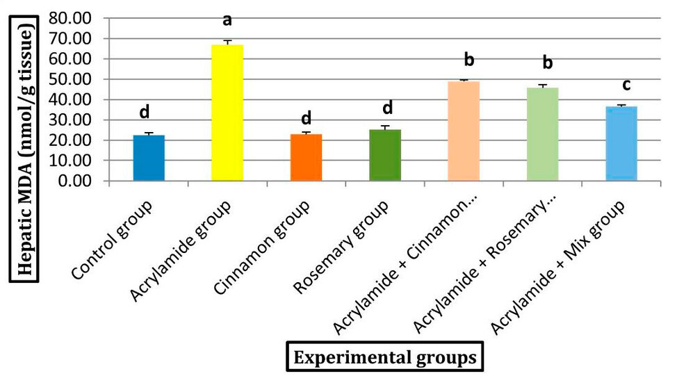
Figure 1: Effect of acrylamide, Cinnamon, and Rosemary oil on hepatic MDA.
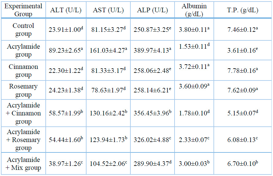
Data are presented as (Mean ± S.E.). S.E. = Standard error.
Mean values with different superscript letters in the same column significantly differ at (P<0.05).
Table 1: Effect of acrylamide, Cinnamon, and rosemary oil on serum ALT, AST, ALP, Albumin, and T.P.
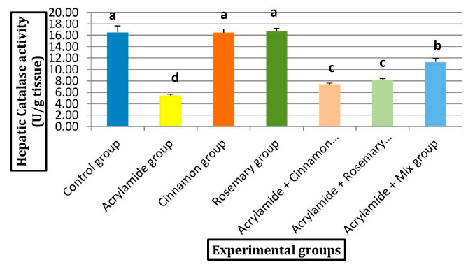
Figure 2: Effect of acrylamide, Cinnamon, and Rosemary oil on Hepatic catalase activity.
Table (2) displays the data as Mean ± S.E., where S.E. represents the standard error. The mean values in the same column with distinct superscript letters indicate significant differences below 0.05 (P<0.05).
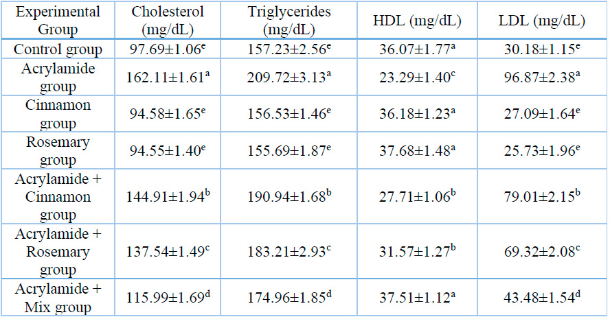
Table 2: Effect of acrylamide, Cinnamon, and Rosemary oil on lipid profile.
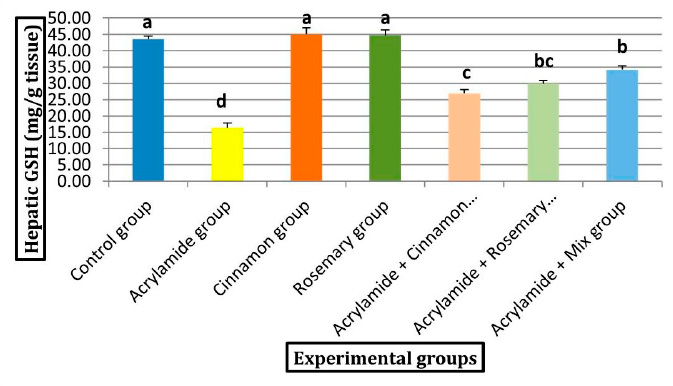
Figure 3: Effect of acrylamide, Cinnamon, and Rosemary oil on Hepatic GSH.
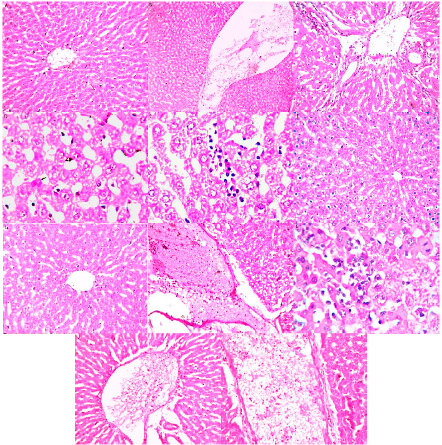
Figure 4: Histopathological examination findings of the hepatic tissue.
The histopathological examination of the hepatic tissue in the control group (A) revealed no histopathological changes. The histological structure of the central vein and the hepatocytes around it within the lobules of the parenchyma appeared normal. The acrylamide group showed (B) thrombosis in the lumen of the dilated central vein and (C) associated with edema and a few inflammatory cell infiltrations in the portal area, as well as dilatation in the portal vein as well as in the bile ducts. (D) There was focal inflammatory cell infiltration and (E) diffuse Kupffer cell proliferation between the degenerated hepatocytes. The cinnamon group showed (F) no histopathological alteration. The rosemary group showed (G) no histopathological alteration. The ACR+Cin group showed (H) thrombosis in the lumen of the dilated portal vein (I) associated with focal inflammatory cell infiltration between the hepatocytes. The ACR+ Ros group showed (J) thrombosis in the lumen of the dilated portal vein. ACR+Cin+Ros showed (K) thrombosis in the lumen of the dilated portal vein.
DISCUSSION
The liver's crucial role in metabolism makes it prone to damage caused by exposure to chemicals. Chemical-induced liver injury poses a significant health risk, often leading to acute liver failure 16. The primary reason behind this specific liver damage is the generation of reactive oxygen species. These reactive compounds cause oxidative stress, subsequently damaging cellular molecules. Consequently, oxidative stress is a well-established contributor to the onset of diverse liver diseases 17.
Oxidative stress plays a significant role in the process of tissue damage. It occurs due to an imbalance between the production of reactive oxygen species and the body's ability to neutralize or counteract their harmful effects 18.
The use of natural antioxidant products has been rapidly growing globally, owing to their proven efficacy and safety. These products are increasingly employed in treating various liver ailments, reflecting their rising popularity in the medical field 3.
Acrylamide is a byproduct formed when starchy foods like potatoes and bread are cooked at high temperatures above 120°C, such as frying or baking. Acrylamide harms living organisms, including genotoxic, neurotoxic, carcinogenic, nephrotoxic, and hepatotoxic 19.
The production of free radicals by acrylamide leads to liver damage. Free radicals are highly reactive and can severely damage cell membranes, nucleic acid, and proteins 20.
Abdel-Daim found that acrylamide has dangerous effects on liver and kidney tissues by increasing the occurrence of lipid peroxidation and altering antioxidant enzyme systems. The significant increase in lipid peroxidation in the liver and kidney can affect the oxidative stress levels in an organism 21.
In this study, we provided evidence that co-treatment with Cinnamon and Rosemary can improve liver function, histology, and oxidative stress caused by acrylamide exposure. This study showed that oral administration of acrylamide at a dose of 20 mg/kg body weight for four weeks led to a significant increase in liver enzymes and cholesterol, triglycerides, and hepatic MDA levels. There was also a significant decrease in albumin, T.P., HDL, and hepatic CAT and GSH levels compared to other groups.
These findings align with a previous study by Erfan 22, who reported elevated levels of liver enzymes ALT and AST and evident histopathological liver injury in the group treated with acrylamide.
The current study showed that the acrylamide-treated group had significantly higher levels of MDA in liver tissue, indicating severe oxidative damage. These findings are consistent with previous studies 5, 23 that also observed increased levels of AST and ALT due to acrylamide administration.
A study conducted by Elhelaly 23 found that oral administration of acrylamide significantly increased levels of ALT and AST, damaging tissue membranes. Acrylamide impaired the viability of hepatic tissue and caused acute toxic damage over time. They also indicated that acrylamide oral administration significantly increased MDA levels and decreased GPX and SOD levels. 23
The present study demonstrated a significant increase in total cholesterol (T.C.) and triglycerides (T.G.); this comes in agreement with Akram24, who reported that low doses of acrylamide caused elevated T.C. and T.G. levels compared to the control group due to lipid peroxidation.
On the other hand, co-treatment with rosemary extract reduced elevated levels of ALT, AST, and ALP in the serum and improved the oxidant-antioxidant status, as seen by decreased MDA levels and increased GSH levels 23, 25.
These results align with the findings of Samaha 26, who reported that carnosic acid prevented liver damage and disturbances in lipid metabolism, leading to decreased levels of ALT, AST, and ALP in the serum.
In another study conducted by Raskovic et al. 27, it was observed that liver injury led to increased levels of AST and ALT enzymes due to hepatocyte damage and leakage. However, treatment with Rosemary was found to mitigate these effects, as indicated by the reduction in elevated MDA levels, which signify oxidative damage to cell membranes. The researchers concluded that adding rosemary supplements partially restored the altered biochemical parameters and significantly reversed markers associated with oxidative stress in experimental models of liver injury.
A study done by Zhao et al. 28 discovered that adding Rosemary to the diet of mice fed a high-fat diet resulted in significant reductions in body weight gain, fat percentage, plasma ALT, triglycerides, free fatty acids, and levels of MDA in the liver and plasma. Furthermore, Rosemary improved liver histology and function, as indicated by a significant decrease in serum levels of ALT and AST, which are markers of liver injury.
Oxidative stress is countered by the complex antioxidant system in mammals. MDA, a byproduct of lipid peroxidation, indicates the level of oxidative damage in the liver tissue.
Cinnamon, a perennial tropical herb from the Lauraceae family, contains essential oil, cinnamaldehyde, cinnamic acid, and cinnamate. Its medicinal properties, including antioxidant, antidiabetic, anti-inflammatory, cardiovascular, anti-cancer, and lipid-lowering effects, make it helpful in treating inflammatory conditions. Cinnamon can reduce organ damage by alleviating oxidative stress 29. Moreover, Cinnamon has protective properties that guard against liver abnormalities in rats.
The current study showed a significant decrease in average ALT and AST levels and a slight reduction in T.C. and T.G. in the livers of rats treated with Cinnamon, Rosemary, or both. These findings align with Ismail et al. 30, who observed significant decreases in ALT and AST, triglycerides, and cholesterol levels in rats with liver damage upon cinnamon administration.
Shalaby and Saifan 31 discovered that the use of cinnamon aqueous extract in diabetic obese rats resulted in a reduction in lipid levels, and it induced liver protection owing to its antioxidant properties. The hepatoprotective effect of cinnamon extract aids in accelerating the recovery and growth of liver cells by promoting the synthesis of proteins. It stimulates the enzyme RNA polymerase 1, which increases ribosomal protein production and facilitates the regeneration of damaged hepatocytes 32.
The consumption of Cinnamon in the diet inhibits the activity of hydroxy-3-methylglutaryl-coenzyme A [HMG Co-A] reductase in the liver, which leads to a decrease in lipids and improved lipid peroxidation. This is achieved by enhancing the functioning of lipid antioxidant enzymes. Additionally, Cinnamon helps in the expression of the LDL-R gene, which contributes to its lipid-lowering effects 33.
Pre-treatment with 200mg/kg/day of cinnamon aqueous extract for 14 days before administering a single toxic dose of acetaminophen significantly reduced the elevated level of ALT and AST levels 34. This provides evidence for the hepatoprotective nature of Cinnamon and its capability to repair liver damage. The positive effects of Cinnamon on liver damage may be attributed to its ability to enhance the antioxidant defense system, scavenge reactive oxygen species, and inhibit lipid peroxidation.
Studies have revealed that cinnamon oil benefits liver toxicity, lipid oxidation, inflammation (specifically IL-1β, IL-6), DNA fragmentation, and histopathological alterations caused by acetaminophen 35. The potential protective effects of Cinnamon can be attributed to its polyphenols and flavonoids, which act as scavengers for reactive oxygen and nitrogen species, chelators for redox-active transition metals, and modulators of enzymes.
Shatwan et al. 36 observed that Cinnamon and ginger decreased the high levels of total cholesterol (T.C.) and triglycerides (T.G.) in men and rats.
The demonstrations by Hindawy and Hendawy 37 were biochemical and histopathological alterations caused by acrylamide on rat livers, and the administration of the potent antioxidant lycopene effectively treated the oxidative damage.
The research by Gawesh et al. 38 discovered that cinnamon administration to rats with liver toxicity significantly reduced the levels of alanine transaminase (ALT), aspartate transaminase (AST), malondialdehyde (MDA), and tumor necrosis factor (TNF), while simultaneously increasing the levels of superoxide dismutase, glutathione peroxidase, and total antioxidant capacity. They also concluded that Cinnamon and ginger are protective against abnormalities in rats with liver toxicity.
In this study, administering Cinnamon and Rosemary, individually or in combination, to rats with liver toxicity resulted in a significant reduction in MDA levels and a significant elevation in hepatic glutathione (GSH). These findings align with Qin et al. 39, who reported that cinnamon extract supplementation decreased tumor necrosis factor Alpha (TNF-α) and MDA. Dehghan et al. 40 asserted that cinnamon administration led to a significant reduction in MDA and TNF levels, along with increased TAC and SOD, attributed to its ability to scavenge free radicals and its high phenol content.
CONCLUSIONS
Our findings provide clear biochemical and histopathological evidence that the administration of cinnamon and rosemary oils to acrylamide-treated rats significantly decreased serum levels of AST and ALT and efficiently reduced hepatic MDA levels; they also increased GSH and CAT activity in liver tissue. Cinnamon and rosemary oils treatment mitigated liver damage associated with acrylamide treatment, attributed to their potent antioxidant activities. We highly recommend using cinnamon and rosemary oils with people with a high frequency of exposure to acrylamide.
Author Contributions: Conceptualization, H.E. and F.E.; methodology, H.E.; investigation, H.E.; resources, E.A.; data curation, H.E.; writing—original draft preparation, H.E.; writing—review and editing, F.E.; visualization, H.E.; supervision, A.E.; project administration, A.E. All authors have read and agreed to the published version of the manuscript.
Funding: This research received no external funding.
Institutional Review Board Statement: The study followed the guidelines in the 8th edition of the National Institutes of Health's Guide for the Care and Use of Laboratory Animals. Additionally, it adhered to the principles set forth by the International Council for Laboratory Animal Science (ICLAS) and those of the Benha University Animal Care and Use Committee, assigned with the identification number (03-03-23).
Informed Consent Statement: Not applicable.
Data Availability Statement: Data is available on request.
Acknowledgments: No acknowledgment to be mentioned.
Conflicts of Interest: The authors declare no conflict of interest.
REFERENCES
1. Savary, S. et al. The global burden of pathogens and pests on major food crops. Nat. Ecol. Evol. 3, (2019).
2. Castillo, J. Identificación de especies de Meloidogyne spp. presentes en el municipio de Patzicía, Chimaltenango. (Universidad Rafael Landívar, 2014).
3. Sánchez-Moreno, S. & Talavera, M. Los nematodos como indicadores ambientales en agroecosistemas. Ecosistemas 22, (2013).
4. Pérez-Anzúrez, G. et al. Arthrobotrys musiformis (Orbiliales) Kills Haemonchus contortus Infective Larvae (Trichostronylidae) through Its Predatory Activity and Its Fungal Culture Filtrates. Pathogens 11, (2022).
5. Triviño Gilces, C., Navia Santillán, D. & Velasco Velasco, L. Guía para reconocer daño en raíces y métodos de muestreo y extracción de nemátodos en raíces y suelo. INIAP Boletín Divulgativo No. 433 https://repositorio.iniap.gob.ec/bitstream/41000/3849/1/433.PDF (2013).
6. González Cardona, C. & Aristizabal Loaiza, M. Evaluación de un producto nematicida sobre nematodos fitoparásitos del plátano Dominico Hartón (Musa AAB). Acta Agron. 63, 71–79 (2014).
7. López-Alcántara, R. Nematodos, su implicación en la producción agrícola. ECUADOR ES Calid. Rev. Científica Ecuatoriana 2, 10–11 (2015).
8. Muthee Gakuubi, M., Wanzala, W., Wagacha, J. M. & Dossaji, S. F. Bioactive properties of Tagetes minuta L. (Asteraceae) essential oils: A review. Am. J. Essent. Oils Nat. Prod. 4, 27–36 (2016).
9. Ibrahim, S. K., Traboulsi, A. F. & El-Haj, S. View of Effects of Essential Oils and Plant Extracts on Hatching, Migration and Mortality of Meloidogyne incognita | Phytopathologia Mediterranea. Phytopathol. Mediterr. 45, 238–246 (2006).
10. Licet Mena Valdés, L. et al. Determinación de saponinas y otros metabolitos secundarios en extractos acuosos de Sapindus saponaria L. (jaboncillo). Rev. Cuba. Plantas Med. 20, 106–116 (2015).
11. Piska, K., Ziaja, K. & Muszynska, B. Edible mushroom pleurotus ostreatus (Oyster mushroom) – Its dietary significance and biological activity. Acta Sci. Pol. Hortorum Cultus 16, 151–161 (2017).
12. Arteaga, M. B., Soria, C. A. & Ordoñez, M. E. Determinación del potencial nematicida y nematostático in vitro de Pleurotus ostreatus (Jacq. ex Fr.) sobre larvas J2 de Globodera pallida (Stone). Rev. Ecuat. Med. Cienc. Biol. 41, 45–50 (2020).
13. Álvarez S., D. E., Botina J., J. A., Ortiz C., A. J. & Botina J., L. L. Evaluación nematicida del aceite esencial de Tagetes zypaquirensis en el manejo del nematodo Meloidogyne spp. Rev. Ciencias Agrícolas 33, 22–33 (2016).
14. Abdel-Rahman, F. H., Alaniz, N. M. & Saleh, M. A. Nematicidal activity of terpenoids. http://dx.doi.org/10.1080/03601234.2012.716686 48, 16–22 (2012).
15. Martinotti, M. D., Castellanos, S. J., González, R., Camargo, A. & Fanzone, M. Efecto nematicida de extractos vegetales sobre Meloidogyne incognita Nematicidal effects of extracts of garlic, grape pomace and olive mill waste, on Meloidogyne incognita, on grapevine cv Chardonnay. Rev. la Fac. Ciencias Agrar. 48, 211–224 (2016).
16. Naim, L. et al. Variation of Pleurotus ostreatus (Jacq. Ex Fr.) P. Kumm. (1871) performance subjected to differentdoses and timings of nano-urea. Saudi J. Biol. Sci. 27, 1573–1579 (2020).
17. Cornelius, W. W. & Wycliffe, W. Chapter 90 - Tagetes (Tagetes minuta) Oils. in Essential Oils in Food Preservation, Flavor and Safety (ed. Preedy, V. R.) 791–802 (Academic Press, 2016). doi:https://doi.org/10.1016/B978-0-12-416641-7.00090-0.
18. Singh, P. Management of Plant-parasitic Nematodes by the Use of Botanicals. J. Plant Physiol. Pathol. 02, (2014).
19. de Lara, on et al. La importancia de los nematodos de vida libre.
20. Guzmán-Piedrahita, O. A., Carolina, C. & López-Nicora, H. D. Physiological interactions of plants with plant-parasitic nematodes: A review. Bol. Cient. del Cent. Museos 24, 190–205 (2020).
21. Silva Olivo, J. C. “Evaluación de la actividad insecticida y/o repelente ‘in vivo’ de extracto acuoso de Artemisia absinthium y aceites esenciales de Tagetes minuta y Tagetes zipaquirensis sobre Lasius niger. (Escuela Superior Politécnica de Chimborazo, 2013).
22. Coyne, D. L., Nicol, J. M., Traducción, C.-C. & Verdejo-Lucas, S. Nematología práctica: Una guía de campo y laboratorio. (International Institute of Tropical Agriculture (IITA), 2007).
23. Jaraba, J. D., Lozano, Z. E. & Suárez Padrón, I. E. Meloidogyne incognita (Kofoid and White, 1919) Chitwood 1949 y Meloidogyne arenaria (Neal 1889) Chitwood 1949: Nematodos de las nudosidades radiculares en guayaba (psidium guajava l.) c.V. Manzana en Monteria, Cordoba. Temas Agrar. ISSN-e 0122-7610, Vol. 8, No. 2, 2003, págs. 15-21 8, 15–21 (2003).
24. Carmona, R. & Padilla, W. Morphological, morphometric and molecular identification of Meloidogyne exigua (Göeldi 1887) in coffee (Coffea arabica). Agron. Mesoam. 31, 531–545 (2020).
25. ICA. Manual para la elaboración de protocolos para ensayos de eficacia con PQUA. (Instituto Colombiano Agropecuario, 2020).
26. Murga-Gutiérrez, S. N., Alvarado-Ibáñez, J. C. & Vera-Obando, N. Y. Efecto del follaje de Tagetes minuta sobre la nodulación radicular de Meloidogyne incognita en Capsicum annuum, en invernadero. Rev. peru. biol 19, 257–260 (2012).
27. Iannacone, J. et al. Acute and chronic toxic effect of Tagetes minuta' Black mint' (Asteraceae) and carbaril on six important entomophages in biological control. Biol. 15, 85–97 (2017).
28. Zygadlo, J. A., Lamarque, A. L., Maestri, D. M., Guzman, C. A. & Grosso, N. R. Composition of the Inflorescence Oils of Some Tagetes Species from Argentina. J. Essent. Oil Res. 5, 679–681 (1993).
29. Peralta-Sánchez, M. G. et al. Metabolitos secundarios y clorofilas en cempasúchil en respuesta a estrés salino. Rev. Mex. ciencias agrícolas 5, 1589–1599 (2014).
30. Senatore, F. et al. Antibacterial activity of Tagetes minuta L. (Asteraceae) essential oil with different chemical composition. Flavour Fragr. J. 19, 574–578 (2004).
31. Alejandro Rojas, G. et al. Evaluación in vitro de la actividad nematicida de limoneno, isotiocianato de alilo, eucaliptol, β-citrolenol y azadiractina sobre Meloidogyne incognita (Nematoda, Meloidogynidae). Trop. Subtrop. Agroecosystems 22, (2019).
32. Herrera Moncada, W. L. & Sandoval Fuentes, M. G. Toxicidad del extracto etanólico de plantas de campo y callos in vitro de Tagetes minuta y Tagetes erecta sobre Meloidogyne spp. en Solanum lycopersicum L. Universidad Nacional Pedro Ruiz Gallo (Universidad Nacional Pedro Ruiz Gallo, 2019).
33. Zarate-Escobedo, J. et al. Concentrations and application intervals of the essential oil of Tagetes lucida Cav. against Nacobbus aberrans. Rev. Mex. Ciencias Agrícolas 9,.
34. Mendoza-García, E. et al. Efecto biológico del aceite de Tagetes coronopifolia (Asteraceae) contra Diaphorina citri (Hemiptera: Liviidae). Rev. Colomb. Entomol. 41, 157–162 (2015).
35. Erazo Sandoval, N. S. et al. Effect of Pleurotus ostreatus (Jacq.) and Trichoderma harzianum (Rifai) on Meloidogyne incognita (Kofoid & White) in tomato (Solanum lycopersicum Mill.). Acta Sci. Biol. Sci. 42, (2020).
36. Clémençon, H., Emmett, V. & Emmett, E. E. Cytology and Plectology of the Hymenomycetes. (2012).
37. Armas-Tizapantzi, A. et al. Estructuras tipo toxocistos en Pleurotus ostreatus y P. pulmonarius. Sci. fungorum 49, e1250 (2019).
38. Ernesto, J., El, S., De La, C., Sur, F. & Royse, D. J. La Biología, el cultivo y las propiedades nutricionales y medicinales de las setas Pleurotus spp. Edible mushroom cultivation View project oxidorreductases enzymes View project. (2017).
39. Aguilar Marcelino, L. et al. Los hongos del género Pleurotus como agentes de biocontrol de parásitos de importancia pecuaria. 52, 1375 (2021).
40. Quevedo, A. et al. Interacciones ecológicas de los hongos nematófagos y su potencial uso en cultivos tropicales. Sci. Agropecu. 13, 97–108 (2022).
41. Jansson, H.-B. & Lopez-Llorca, L. V. Hongos nematófagos. 145–173 https://dcmba.ua.es/es/areas/botanica/hongos-nematofagos.html# (2001).
42. Leonardo, H. et al. Activity of the fungus Pleurotus ostreatus and of its proteases on Panagrellus sp. larvae. African J. Biotechnol. 14, 1496–1503 (2015).
43. Arteaga Paredes, M. B. Determinación del potencial nematicida y nematostático in vitro de Pleurotus ostreatus (Agaricales: Pleurotaceae) sobre larvas J2 de Globodera pallida (Tylenchida: Heteroderidae). (Pontificia Universidad Católica del Ecuador, 2018).
Received: October 9th 2023/ Accepted: January 15th 2024 / Published:15 February 2024
Citation: Elsayed H., El komy A. A. E., El-Shewy E. A., Abdallah F. E. E. Ameliorative Effect of Cinnamon and Rosemary Oils in Acrylamide–Induced Hepatic Injury in Rats. Revis Bionatura 2024; 1 (1) 58. http://dx.doi.org/10.21931/BJ/2024.01.01.58
Additional information. Correspondence should be addressed to [email protected].
Peer review information. Bionatura thanks anonymous reviewer(s) for their contribution to the peer review of this work using https://reviewerlocator.webofscience.com/
All articles published by Bionatura Journal are made freely and permanently accessible online immediately upon publication, without subscription charges or registration barriers.
Publisher's Note: Bionatura stays neutral concerning jurisdictional claims in published maps and institutional affiliations.
Copyright: © 2024 by the authors. They were submitted for possible open-access publication under the terms and conditions of the Creative Commons Attribution (CC BY) license (https://creativecommons.org/licenses/by/4.0/).
